

89
Anatomy
body of the longer pleuronematic flagellum. After leaving the basal body, this fibrous root passes
closely against the edge of the chloroplast and reaches the tip of the pyriform nucleus. It then splits
into several branches, ramifying over the nuclear surface, with some branches located in the narrow
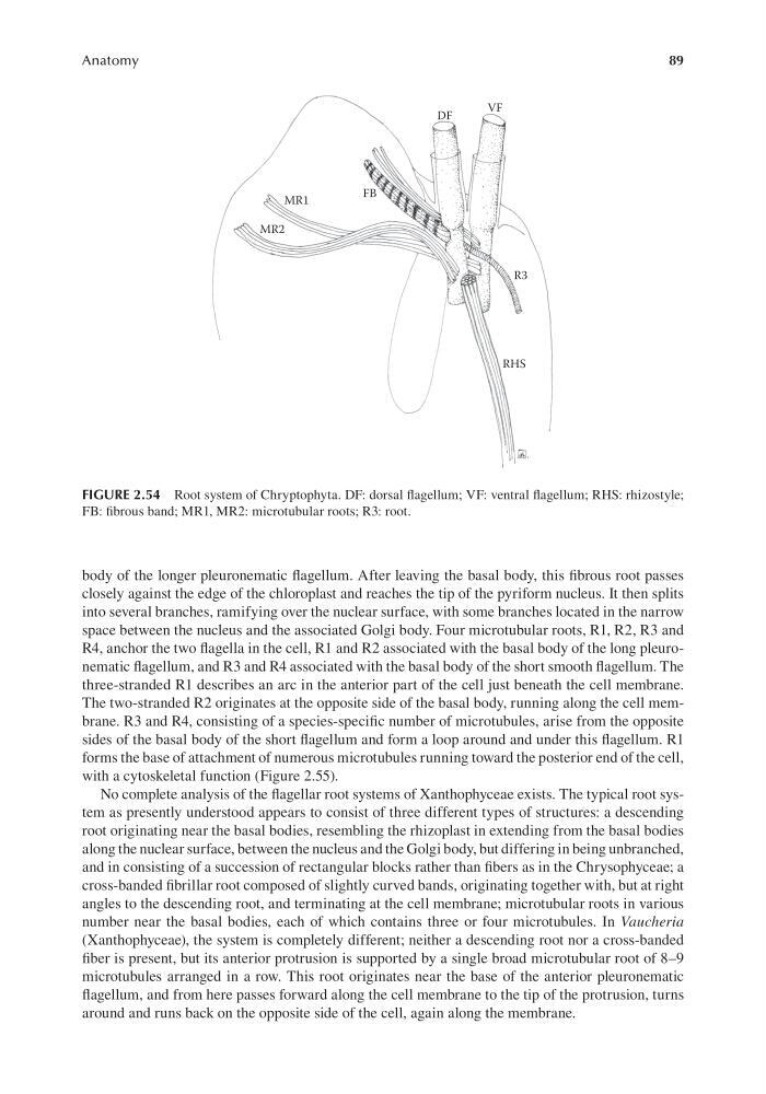
space between the nucleus and the associated Golgi body. Four microtubular roots, R1, R2, R3 and
R4, anchor the two flagella in the cell, R1 and R2 associated with the basal body of the long pleuronematic flagellum, and R3 and R4 associated with the basal body of the short smooth flagellum. The
three-stranded R1 describes an arc in the anterior part of the cell just beneath the cell membrane.
The two-stranded R2 originates at the opposite side of the basal body, running along the cell membrane. R3 and R4, consisting of a species-specific number of microtubules, arise from the opposite
sides of the basal body of the short flagellum and form a loop around and under this flagellum. R1
forms the base of attachment of numerous microtubules running toward the posterior end of the cell,
with a cytoskeletal function (Figure 2.55).
No complete analysis of the flagellar root systems of Xanthophyceae exists. The typical root system as presently understood appears to consist of three different types of structures: a descending
root originating near the basal bodies, resembling the rhizoplast in extending from the basal bodies
along the nuclear surface, between the nucleus and the Golgi body, but differing in being unbranched,
and in consisting of a succession of rectangular blocks rather than fibers as in the Chrysophyceae; a
cross-banded fibrillar root composed of slightly curved bands, originating together with, but at right
angles to the descending root, and terminating at the cell membrane; microtubular roots in various
number near the basal bodies, each of which contains three or four microtubules. In Vaucheria
(Xanthophyceae), the system is completely different; neither a descending root nor a cross-banded
fiber is present, but its anterior protrusion is supported by a single broad microtubular root of 8–9
microtubules arranged in a row. This root originates near the base of the anterior pleuronematic
flagellum, and from here passes forward along the cell membrane to the tip of the protrusion, turns
around and runs back on the opposite side of the cell, again along the membrane.
DF
VF
FB
R3
RHS
MR1
MR2
FIGURE 2.54 Root system of Chryptophyta. DF: dorsal flagellum; VF: ventral flagellum; RHS: rhizostyle;
FB: fibrous band; MR1, MR2: microtubular roots; R3: root.
Anatomy
body of the longer pleuronematic flagellum. After leaving the basal body, this fibrous root passes
closely against the edge of the chloroplast and reaches the tip of the pyriform nucleus. It then splits
into several branches, ramifying over the nuclear surface, with some branches located in the narrow
space between the nucleus and the associated Golgi body. Four microtubular roots, R1, R2, R3 and
R4, anchor the two flagella in the cell, R1 and R2 associated with the basal body of the long pleuronematic flagellum, and R3 and R4 associated with the basal body of the short smooth flagellum. The
three-stranded R1 describes an arc in the anterior part of the cell just beneath the cell membrane.
The two-stranded R2 originates at the opposite side of the basal body, running along the cell membrane. R3 and R4, consisting of a species-specific number of microtubules, arise from the opposite
sides of the basal body of the short flagellum and form a loop around and under this flagellum. R1
forms the base of attachment of numerous microtubules running toward the posterior end of the cell,
with a cytoskeletal function (Figure 2.55).
No complete analysis of the flagellar root systems of Xanthophyceae exists. The typical root system as presently understood appears to consist of three different types of structures: a descending
root originating near the basal bodies, resembling the rhizoplast in extending from the basal bodies
along the nuclear surface, between the nucleus and the Golgi body, but differing in being unbranched,
and in consisting of a succession of rectangular blocks rather than fibers as in the Chrysophyceae; a
cross-banded fibrillar root composed of slightly curved bands, originating together with, but at right
angles to the descending root, and terminating at the cell membrane; microtubular roots in various
number near the basal bodies, each of which contains three or four microtubules. In Vaucheria
(Xanthophyceae), the system is completely different; neither a descending root nor a cross-banded
fiber is present, but its anterior protrusion is supported by a single broad microtubular root of 8–9
microtubules arranged in a row. This root originates near the base of the anterior pleuronematic
flagellum, and from here passes forward along the cell membrane to the tip of the protrusion, turns
around and runs back on the opposite side of the cell, again along the membrane.
DF
VF
FB
R3
RHS
MR1
MR2
FIGURE 2.54 Root system of Chryptophyta. DF: dorsal flagellum; VF: ventral flagellum; RHS: rhizostyle;
FB: fibrous band; MR1, MR2: microtubular roots; R3: root.