

85
Anatomy
The flagellar apparatus of the reproductive cells of the Bryopsidales (Ulvophyceae) is anchored
in the cell by four microtubular roots following the usual X–2–X–2 pattern. Each of the microtubules in the rootlet may be subtended by an electron-dense wing. The basal bodies are connected
anteriorly by a nonstriated upper connective, with a typically pronounced arched appearance.
Transversely striated bands connecting the rootlets to the basal bodies are also present. Species
exist in this class, which produce stephanokont zoospores with more than 30 flagella. In these cells,
the basal bodies are connected by a nonstriated fibrous upper ring, which can be considered the
result of the fusion of many nonstriated upper connectives. The proximal ends of the basal bodies are partially enclosed in a second lower ring of amorphous material. Four- and six-membered
microtubular roots depart from between the basal bodies. The microtubular root system of the
Trentepohliales (Ulvophyceae) motile cells (tetraflagellate zoospores and biflagellate gametes) does
not follow the X–2–X–2 pattern, but shows a 6–4– 6–4 arrangement in Trentepohlia sp. The dorsoventrally compressed form of the flagellate cells forces the basal bodies and the root system components in a flattened arrangement. Basal bodies are aligned perpendicular to the long axis of the cell,
with a parallel or antiparallel arrangement, and the microtubular rootlets, especially in the gametes,
extend posteriorly nearly parallel to one another and to the basal body from which they arise. Two of
the rootlets (the 4-membered rootlets in Trentepohlia sp.) are associated with a complex columnar
structure resembling the multilayered structure of the Charophyceae.
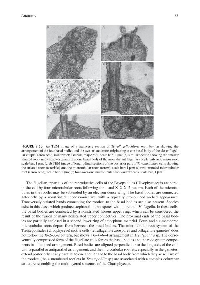
FIGURE 2.50 (a) TEM image of a transverse section of Tetraflagellochloris mauritanica showing the
arrangement of the four basal bodies and the two striated roots originating at one basal body of the closer flagellar couple; arrowhead, minor root; asterisk, major root, scale bar, 1 μm; (b) similar section showing the smaller
striated root (arrowhead) originating at one basal body of the more distant flagellar couple; asterisk, major root,
scale bar, 1 μm; (c, d) TEM image of longitudinal sections of the posterior part of T. mauritanica cells showing
the striated roots (asterisks) and the microtubular roots (arrow), scale bar: 1 μm; (e) two-stranded microtubular
root (arrowhead), scale bar, 1 μm; (f) four-over-one microtubular root (arrowhead), scale bar, 1 μm.
Anatomy
The flagellar apparatus of the reproductive cells of the Bryopsidales (Ulvophyceae) is anchored
in the cell by four microtubular roots following the usual X–2–X–2 pattern. Each of the microtubules in the rootlet may be subtended by an electron-dense wing. The basal bodies are connected
anteriorly by a nonstriated upper connective, with a typically pronounced arched appearance.
Transversely striated bands connecting the rootlets to the basal bodies are also present. Species
exist in this class, which produce stephanokont zoospores with more than 30 flagella. In these cells,
the basal bodies are connected by a nonstriated fibrous upper ring, which can be considered the
result of the fusion of many nonstriated upper connectives. The proximal ends of the basal bodies are partially enclosed in a second lower ring of amorphous material. Four- and six-membered
microtubular roots depart from between the basal bodies. The microtubular root system of the
Trentepohliales (Ulvophyceae) motile cells (tetraflagellate zoospores and biflagellate gametes) does
not follow the X–2–X–2 pattern, but shows a 6–4– 6–4 arrangement in Trentepohlia sp. The dorsoventrally compressed form of the flagellate cells forces the basal bodies and the root system components in a flattened arrangement. Basal bodies are aligned perpendicular to the long axis of the cell,
with a parallel or antiparallel arrangement, and the microtubular rootlets, especially in the gametes,
extend posteriorly nearly parallel to one another and to the basal body from which they arise. Two of
the rootlets (the 4-membered rootlets in Trentepohlia sp.) are associated with a complex columnar
structure resembling the multilayered structure of the Charophyceae.
FIGURE 2.50 (a) TEM image of a transverse section of Tetraflagellochloris mauritanica showing the
arrangement of the four basal bodies and the two striated roots originating at one basal body of the closer flagellar couple; arrowhead, minor root; asterisk, major root, scale bar, 1 μm; (b) similar section showing the smaller
striated root (arrowhead) originating at one basal body of the more distant flagellar couple; asterisk, major root,
scale bar, 1 μm; (c, d) TEM image of longitudinal sections of the posterior part of T. mauritanica cells showing
the striated roots (asterisks) and the microtubular roots (arrow), scale bar: 1 μm; (e) two-stranded microtubular
root (arrowhead), scale bar, 1 μm; (f) four-over-one microtubular root (arrowhead), scale bar, 1 μm.