

84
Algae
four flagella in the cell by a cruciate root system. One pair of striated roots, with one root more
prominent than the other, originates at each of the two closer basal bodies (Figure 2.50a, white
asterisk and white arrowhead, respectively). Smaller striated roots originate at the second pair
of basal bodies (Figure 2.50b, arrowhead). In transverse and longitudinal sections, these striated
roots reveal a periodicity of 300–330 nm (Figure 2.50c, asterisk) and a clear distal tapering where
they underlie the plasmalemma (Figure 2.50c). Microtubular roots are also present: two-stranded
microtubular roots (Figure 2.50e, arrows) and four-over-one roots (Figure 2.50f, arrow). The striated roots are closely associated with the microtubular roots (Figure 2.50c and d, arrow). In TEM
and SEM images (Figure 2.51a and b), the tapered ends of the striated roots form two symmetrical
shoulders in the posterior part of the cell, just below the flagellar emergence; the microtubular
roots curve above the shoulders and down the sides of the cell for some distance. Basal bodies no.
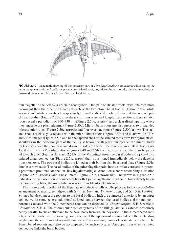
1 and no. 2 lie in a V configuration (Figures 2.49 and 2.51c), while those of the other pair lie parallel to each other (Figures 2.49 and 2.51d). In the V configuration, the basal bodies are joined by a
striated distal connection (Figure 2.51c, arrow) that is positioned immediately below the flagellar
transition zone. The two basal bodies are joined at their bottom also by a basal plate (Figure 2.51c,
double arrowheads). The basal bodies of the other flagellar pair show a similar connection system;
a prominent proximal connection showing alternating electron-dense zones resembling a striation
(Figure 2.51d, asterisk) and a basal plate (Figure 2.51c, arrowheads). The arrow in Figure 2.51d
indicates the cross-sectioned connecting fiber that joins flagella no. 1 and no. 2. Immediately below
the connecting fiber, the microtubular roots are visible (double asterisks).
The microtubular rootlets of the flagellate reproductive cells of Ulvophyceae follow the X–2–X–2
arrangement of most green algae, with X = 4 in Ulva and Enteromorpha, and X = 5 in Ulothrix.
Striated bands connect the rootlets to the basal bodies, which are connected anteriorly by an upper
connective; in some genera, additional striated bands between the basal bodies and striated component associated with the 2-membered root can be detected. In Chaetomorpha, X is 3, while in
Cladophora X is 4. The microtubular rootlet systems of the biflagellate cells extends posteriorly
nearly parallel to one another and to the basal body from which they arise. In the X-membered rootlets, an electron-dense strut or wing connects one of the uppermost microtubules to the subtending
singlet, and the entire rootlet is usually subtended by a massive, more or less striated structure. The
2-membered rootlets may also be accompanied by such structures. An upper transversely striated
connective links the basal bodies.
FIGURE 2.49 Schematic drawing of the posterior part of Tetraflagellochloris mauritanica illustrating the
main components of the flagellar apparatus; sr, striated root; mr, microtubular root; dc, distal connection; pc,
proximal connection; bp, basal plate. See text for details.
Algae
four flagella in the cell by a cruciate root system. One pair of striated roots, with one root more
prominent than the other, originates at each of the two closer basal bodies (Figure 2.50a, white
asterisk and white arrowhead, respectively). Smaller striated roots originate at the second pair
of basal bodies (Figure 2.50b, arrowhead). In transverse and longitudinal sections, these striated
roots reveal a periodicity of 300–330 nm (Figure 2.50c, asterisk) and a clear distal tapering where
they underlie the plasmalemma (Figure 2.50c). Microtubular roots are also present: two-stranded
microtubular roots (Figure 2.50e, arrows) and four-over-one roots (Figure 2.50f, arrow). The striated roots are closely associated with the microtubular roots (Figure 2.50c and d, arrow). In TEM
and SEM images (Figure 2.51a and b), the tapered ends of the striated roots form two symmetrical
shoulders in the posterior part of the cell, just below the flagellar emergence; the microtubular
roots curve above the shoulders and down the sides of the cell for some distance. Basal bodies no.
1 and no. 2 lie in a V configuration (Figures 2.49 and 2.51c), while those of the other pair lie parallel to each other (Figures 2.49 and 2.51d). In the V configuration, the basal bodies are joined by a
striated distal connection (Figure 2.51c, arrow) that is positioned immediately below the flagellar
transition zone. The two basal bodies are joined at their bottom also by a basal plate (Figure 2.51c,
double arrowheads). The basal bodies of the other flagellar pair show a similar connection system;
a prominent proximal connection showing alternating electron-dense zones resembling a striation
(Figure 2.51d, asterisk) and a basal plate (Figure 2.51c, arrowheads). The arrow in Figure 2.51d
indicates the cross-sectioned connecting fiber that joins flagella no. 1 and no. 2. Immediately below
the connecting fiber, the microtubular roots are visible (double asterisks).
The microtubular rootlets of the flagellate reproductive cells of Ulvophyceae follow the X–2–X–2
arrangement of most green algae, with X = 4 in Ulva and Enteromorpha, and X = 5 in Ulothrix.
Striated bands connect the rootlets to the basal bodies, which are connected anteriorly by an upper
connective; in some genera, additional striated bands between the basal bodies and striated component associated with the 2-membered root can be detected. In Chaetomorpha, X is 3, while in
Cladophora X is 4. The microtubular rootlet systems of the biflagellate cells extends posteriorly
nearly parallel to one another and to the basal body from which they arise. In the X-membered rootlets, an electron-dense strut or wing connects one of the uppermost microtubules to the subtending
singlet, and the entire rootlet is usually subtended by a massive, more or less striated structure. The
2-membered rootlets may also be accompanied by such structures. An upper transversely striated
connective links the basal bodies.
FIGURE 2.49 Schematic drawing of the posterior part of Tetraflagellochloris mauritanica illustrating the
main components of the flagellar apparatus; sr, striated root; mr, microtubular root; dc, distal connection; pc,
proximal connection; bp, basal plate. See text for details.