

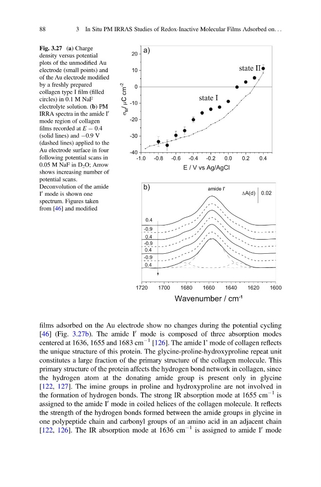
films adsorbed on the Au electrode show no changes during the potential cycling
[46] (Fig. 3.27b). The amide I
0 mode is composed of three absorption modes
centered at 1636, 1655 and 1683 cm
À1 [126]. The amide I’ mode of collagen reflects
the unique structure of this protein. The glycine-proline-hydroxyproline repeat unit
constitutes a large fraction of the primary structure of the collagen molecule. This
primary structure of the protein affects the hydrogen bond network in collagen, since
the hydrogen atom at the donating amide group is present only in glycine
[122, 127]. The imine groups in proline and hydroxyproline are not involved in
the formation of hydrogen bonds. The strong IR absorption mode at 1655 cm
À1 is
assigned to the amide I
0 mode in coiled helices of the collagen molecule. It reflects
the strength of the hydrogen bonds formed between the amide groups in glycine in
one polypeptide chain and carbonyl groups of an amino acid in an adjacent chain
[122, 126]. The IR absorption mode at 1636 cm
À1 is assigned to amide I
0 mode
-1.0
-0.8
-0.6
-0.4
-0.2
0.0
0.2
0.4
-40
-30
-20
-10
0
10
20
state II
M /
m
c
C
-2
E / V vs Ag/AgCl
a)
state I
1720
1700
1680
1660
1640
1620
1600
-0.9
-0.9
0.4
0.4
0.4
-0.9
amide I'
Wavenumber / cm
-1
A(d) 0.02
0.4
b)
Fig. 3.27 (a) Charge
density versus potential
plots of the unmodified Au
electrode (small points) and
of the Au electrode modified
by a freshly prepared
collagen type I film (filled
circles) in 0.1 M NaF
electrolyte solution. (b) PM
IRRA spectra in the amide I
0
mode region of collagen
films recorded at E ¼ 0.4
(solid lines) and À0.9 V
(dashed lines) applied to the
Au electrode surface in four
following potential scans in
0.05 M NaF in D 2 O; Arrow
shows increasing number of
potential scans.
Deconvolution of the amide
I
0 mode is shown one
spectrum. Figures taken
from [46] and modified
88
3 In Situ PM IRRAS Studies of Redox-Inactive Molecular Films Adsorbed on. . .
[46] (Fig. 3.27b). The amide I
0 mode is composed of three absorption modes
centered at 1636, 1655 and 1683 cm
À1 [126]. The amide I’ mode of collagen reflects
the unique structure of this protein. The glycine-proline-hydroxyproline repeat unit
constitutes a large fraction of the primary structure of the collagen molecule. This
primary structure of the protein affects the hydrogen bond network in collagen, since
the hydrogen atom at the donating amide group is present only in glycine
[122, 127]. The imine groups in proline and hydroxyproline are not involved in
the formation of hydrogen bonds. The strong IR absorption mode at 1655 cm
À1 is
assigned to the amide I
0 mode in coiled helices of the collagen molecule. It reflects
the strength of the hydrogen bonds formed between the amide groups in glycine in
one polypeptide chain and carbonyl groups of an amino acid in an adjacent chain
[122, 126]. The IR absorption mode at 1636 cm
À1 is assigned to amide I
0 mode
-1.0
-0.8
-0.6
-0.4
-0.2
0.0
0.2
0.4
-40
-30
-20
-10
0
10
20
state II
M /
m
c
C
-2
E / V vs Ag/AgCl
a)
state I
1720
1700
1680
1660
1640
1620
1600
-0.9
-0.9
0.4
0.4
0.4
-0.9
amide I'
Wavenumber / cm
-1
A(d) 0.02
0.4
b)
Fig. 3.27 (a) Charge
density versus potential
plots of the unmodified Au
electrode (small points) and
of the Au electrode modified
by a freshly prepared
collagen type I film (filled
circles) in 0.1 M NaF
electrolyte solution. (b) PM
IRRA spectra in the amide I
0
mode region of collagen
films recorded at E ¼ 0.4
(solid lines) and À0.9 V
(dashed lines) applied to the
Au electrode surface in four
following potential scans in
0.05 M NaF in D 2 O; Arrow
shows increasing number of
potential scans.
Deconvolution of the amide
I
0 mode is shown one
spectrum. Figures taken
from [46] and modified
88
3 In Situ PM IRRAS Studies of Redox-Inactive Molecular Films Adsorbed on. . .