

that the amide groups in the protein make hydrogen bonds to the ester carbonyl
groups in DMPC molecules [35]. Basic amino acids (lysine and arginine) are present
at the N-terminus (K5, K11, K22, K37, R48, K84) of recoverin, which is known to
interact via charge-charge and charge-dipole interactions with lipids present in
membranes [45]. The charge-dipole interactions appear to be stronger with n-Rv
than with Rv, suggesting distinct conformational changes in membrane bound n-Rv
and Rv.
The order parameter of the carbonyl ester group (S (C¼O) ) differs for pure DMPC:
cholesterol bilayer and membrane interacting with n-Rv and Rv [45]. In the DMPC:
cholesterol bilayer adsorbed on the Au electrode surface S (C¼O) ¼ À0.14 Æ 0.05.
During the membrane desorption from the Au surface, S (C¼O) decreases to À0.25,
reflecting changes in orientation of the hydrocarbon chains. In the membrane
with bound n-Rv and Rv S (C¼O) is close to À0.25 and À0.40, respectively. The
1800
1700
1600
1500
1400
S
Wavenumber / cm
-1
c)
(CH 2 )+
as
(CH 3 )+
Amide II'+
s
(COO
-
)
as
(COO-)
(C=O) Lipid
S
(C=O) Lipid
Amide I'
b)
(CH 2 )+
as
(CH 3 )
(NO)
(C=O) Lipid
S
S 0.001
a)
-E
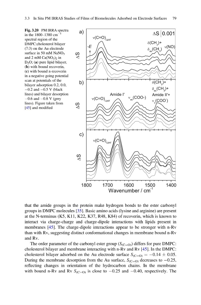
Fig. 3.20 PM IRRA spectra
in the 1800–1380 cm
À1
spectral region of the
DMPC:cholesterol bilayer
(7:3) on the Au electrode
surface in 50 mM NaNO 3
and 2 mM Ca(NO 3 ) 2 in
D 2 O; (a) pure lipid bilayer,
(b) with bound recoverin,
(c) with bound n-recoverin
in a negative going potential
scan at potentials of the
bilayer adsorption 0.2, 0.0,
À0.2 and À0.5 V (black
lines) and bilayer desorption
À0.6 and À0.8 V (grey
lines). Figure taken from
[45] and modified
3.3 In Situ PM IRRAS Studies of Films of Biomolecules Adsorbed on Electrode Surfaces 79
groups in DMPC molecules [35]. Basic amino acids (lysine and arginine) are present
at the N-terminus (K5, K11, K22, K37, R48, K84) of recoverin, which is known to
interact via charge-charge and charge-dipole interactions with lipids present in
membranes [45]. The charge-dipole interactions appear to be stronger with n-Rv
than with Rv, suggesting distinct conformational changes in membrane bound n-Rv
and Rv.
The order parameter of the carbonyl ester group (S (C¼O) ) differs for pure DMPC:
cholesterol bilayer and membrane interacting with n-Rv and Rv [45]. In the DMPC:
cholesterol bilayer adsorbed on the Au electrode surface S (C¼O) ¼ À0.14 Æ 0.05.
During the membrane desorption from the Au surface, S (C¼O) decreases to À0.25,
reflecting changes in orientation of the hydrocarbon chains. In the membrane
with bound n-Rv and Rv S (C¼O) is close to À0.25 and À0.40, respectively. The
1800
1700
1600
1500
1400
S
Wavenumber / cm
-1
c)
(CH 2 )+
as
(CH 3 )+
Amide II'+
s
(COO
-
)
as
(COO-)
(C=O) Lipid
S
(C=O) Lipid
Amide I'
b)
(CH 2 )+
as
(CH 3 )
(NO)
(C=O) Lipid
S
S 0.001
a)
-E
Fig. 3.20 PM IRRA spectra
in the 1800–1380 cm
À1
spectral region of the
DMPC:cholesterol bilayer
(7:3) on the Au electrode
surface in 50 mM NaNO 3
and 2 mM Ca(NO 3 ) 2 in
D 2 O; (a) pure lipid bilayer,
(b) with bound recoverin,
(c) with bound n-recoverin
in a negative going potential
scan at potentials of the
bilayer adsorption 0.2, 0.0,
À0.2 and À0.5 V (black
lines) and bilayer desorption
À0.6 and À0.8 V (grey
lines). Figure taken from
[45] and modified
3.3 In Situ PM IRRAS Studies of Films of Biomolecules Adsorbed on Electrode Surfaces 79