

interaction of recoverin with phospholipids reduces the motional freedom of the
ester group, which is associated with the appearance of electrostatic charge-dipole
interactions between recoverin and the ester carbonyl groups in DMPC molecules.
In the PM IRRA spectra of the lipid membrane interacting with recoverins a
broad IR absorption mode in the 1700–1600 cm
À1 region is observed (Fig. 3.20b,c).
This mode arises from the amide I
0 mode in recoverins bound to the DMPC:
cholesterol bilayer. The amide I
0 mode in the membrane with bound n-Rv has
ca. 3 times larger intensity than that of Rv. This result indicates that a larger amount
of n-Rv than Rv binds to the membrane surface. The maximum of absorption of the
amide I
0 mode is equal to 1648 cm
À1 . Its shape is asymmetric. Deconvolution of the
amide I
0 mode gives three modes centered at 1640–1650, 1666 and 1681 cm
À1 ,
originating from α-helical, disordered and β-turns structural elements [45]. In the
membrane bound state α-helices and disordered or flexible fragments contribute to
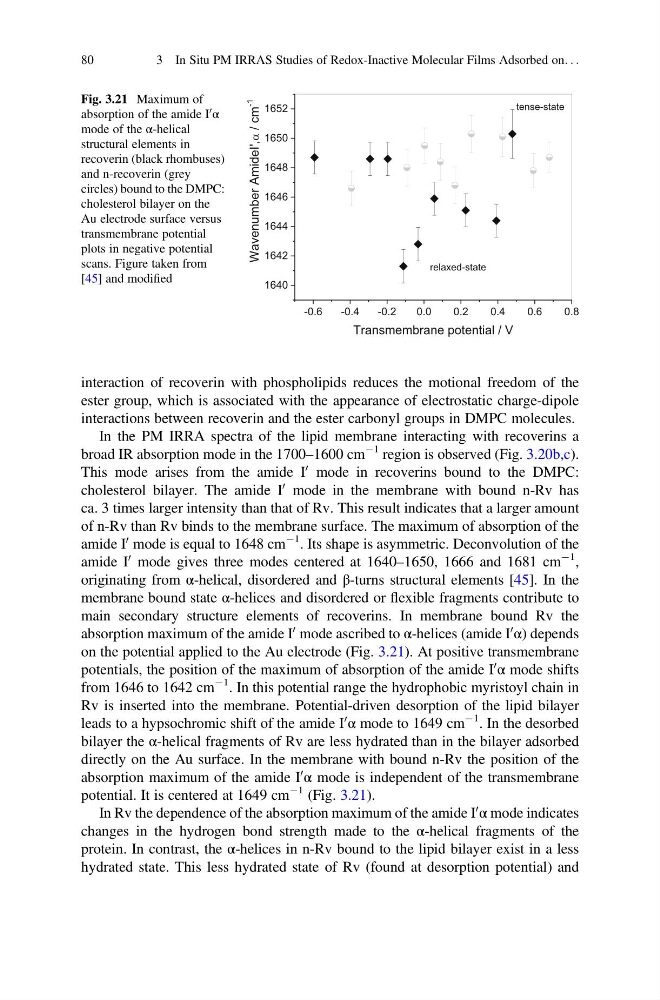
main secondary structure elements of recoverins. In membrane bound Rv the
absorption maximum of the amide I
0 mode ascribed to α-helices (amide I
0
α) depends
on the potential applied to the Au electrode (Fig. 3.21). At positive transmembrane
potentials, the position of the maximum of absorption of the amide I
0
α mode shifts
from 1646 to 1642 cm
À1 . In this potential range the hydrophobic myristoyl chain in
Rv is inserted into the membrane. Potential-driven desorption of the lipid bilayer
leads to a hypsochromic shift of the amide I
0
α mode to 1649 cm
À1 . In the desorbed
bilayer the α-helical fragments of Rv are less hydrated than in the bilayer adsorbed
directly on the Au surface. In the membrane with bound n-Rv the position of the
absorption maximum of the amide I
0
α mode is independent of the transmembrane
potential. It is centered at 1649 cm
À1 (Fig. 3.21).
In Rv the dependence of the absorption maximum of the amide I
0
α mode indicates
changes in the hydrogen bond strength made to the α-helical fragments of the
protein. In contrast, the α-helices in n-Rv bound to the lipid bilayer exist in a less
hydrated state. This less hydrated state of Rv (found at desorption potential) and
-0.6
-0.4
-0.2
0.0
0.2
0.4
0.6
0.8
1640
1642
1644
1646
1648
1650
1652
,
'
I
e
d
i
m
A
r
e
b
m
u
n
e
v
a
W
m
c
/
-1
Transmembrane potential / V
tense-state
relaxed-state
Fig. 3.21 Maximum of
absorption of the amide I
0 α
mode of the α-helical
structural elements in
recoverin (black rhombuses)
and n-recoverin (grey
circles) bound to the DMPC:
cholesterol bilayer on the
Au electrode surface versus
transmembrane potential
plots in negative potential
scans. Figure taken from
[45] and modified
80
3 In Situ PM IRRAS Studies of Redox-Inactive Molecular Films Adsorbed on. . .
ester group, which is associated with the appearance of electrostatic charge-dipole
interactions between recoverin and the ester carbonyl groups in DMPC molecules.
In the PM IRRA spectra of the lipid membrane interacting with recoverins a
broad IR absorption mode in the 1700–1600 cm
À1 region is observed (Fig. 3.20b,c).
This mode arises from the amide I
0 mode in recoverins bound to the DMPC:
cholesterol bilayer. The amide I
0 mode in the membrane with bound n-Rv has
ca. 3 times larger intensity than that of Rv. This result indicates that a larger amount
of n-Rv than Rv binds to the membrane surface. The maximum of absorption of the
amide I
0 mode is equal to 1648 cm
À1 . Its shape is asymmetric. Deconvolution of the
amide I
0 mode gives three modes centered at 1640–1650, 1666 and 1681 cm
À1 ,
originating from α-helical, disordered and β-turns structural elements [45]. In the
membrane bound state α-helices and disordered or flexible fragments contribute to
main secondary structure elements of recoverins. In membrane bound Rv the
absorption maximum of the amide I
0 mode ascribed to α-helices (amide I
0
α) depends
on the potential applied to the Au electrode (Fig. 3.21). At positive transmembrane
potentials, the position of the maximum of absorption of the amide I
0
α mode shifts
from 1646 to 1642 cm
À1 . In this potential range the hydrophobic myristoyl chain in
Rv is inserted into the membrane. Potential-driven desorption of the lipid bilayer
leads to a hypsochromic shift of the amide I
0
α mode to 1649 cm
À1 . In the desorbed
bilayer the α-helical fragments of Rv are less hydrated than in the bilayer adsorbed
directly on the Au surface. In the membrane with bound n-Rv the position of the
absorption maximum of the amide I
0
α mode is independent of the transmembrane
potential. It is centered at 1649 cm
À1 (Fig. 3.21).
In Rv the dependence of the absorption maximum of the amide I
0
α mode indicates
changes in the hydrogen bond strength made to the α-helical fragments of the
protein. In contrast, the α-helices in n-Rv bound to the lipid bilayer exist in a less
hydrated state. This less hydrated state of Rv (found at desorption potential) and
-0.6
-0.4
-0.2
0.0
0.2
0.4
0.6
0.8
1640
1642
1644
1646
1648
1650
1652
,
'
I
e
d
i
m
A
r
e
b
m
u
n
e
v
a
W
m
c
/
-1
Transmembrane potential / V
tense-state
relaxed-state
Fig. 3.21 Maximum of
absorption of the amide I
0 α
mode of the α-helical
structural elements in
recoverin (black rhombuses)
and n-recoverin (grey
circles) bound to the DMPC:
cholesterol bilayer on the
Au electrode surface versus
transmembrane potential
plots in negative potential
scans. Figure taken from
[45] and modified
80
3 In Situ PM IRRAS Studies of Redox-Inactive Molecular Films Adsorbed on. . .