

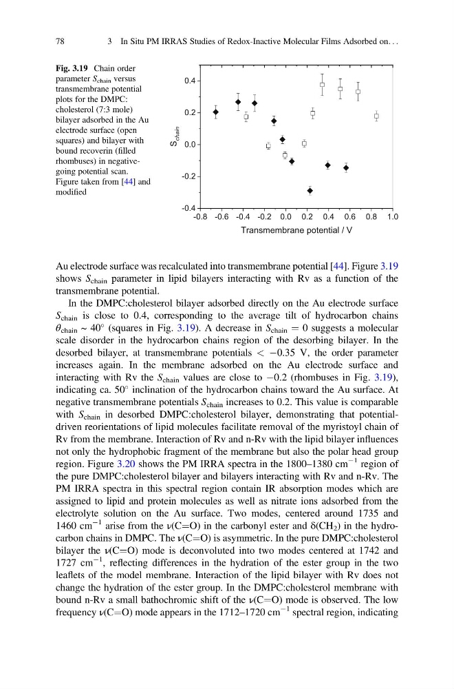
Au electrode surface was recalculated into transmembrane potential [44]. Figure 3.19
shows S chain parameter in lipid bilayers interacting with Rv as a function of the
transmembrane potential.
In the DMPC:cholesterol bilayer adsorbed directly on the Au electrode surface
S chain is close to 0.4, corresponding to the average tilt of hydrocarbon chains
θ chain ~ 40
(squares in Fig. 3.19). A decrease in S chain ¼ 0 suggests a molecular
scale disorder in the hydrocarbon chains region of the desorbing bilayer. In the
desorbed bilayer, at transmembrane potentials < À0.35 V, the order parameter
increases again. In the membrane adsorbed on the Au electrode surface and
interacting with Rv the S chain values are close to À0.2 (rhombuses in Fig. 3.19),
indicating ca. 50
inclination of the hydrocarbon chains toward the Au surface. At
negative transmembrane potentials S chain increases to 0.2. This value is comparable
with S chain in desorbed DMPC:cholesterol bilayer, demonstrating that potentialdriven reorientations of lipid molecules facilitate removal of the myristoyl chain of
Rv from the membrane. Interaction of Rv and n-Rv with the lipid bilayer influences
not only the hydrophobic fragment of the membrane but also the polar head group
region. Figure 3.20 shows the PM IRRA spectra in the 1800–1380 cm
À1 region of
the pure DMPC:cholesterol bilayer and bilayers interacting with Rv and n-Rv. The
PM IRRA spectra in this spectral region contain IR absorption modes which are
assigned to lipid and protein molecules as well as nitrate ions adsorbed from the
electrolyte solution on the Au surface. Two modes, centered around 1735 and
1460 cm
À1 arise from the ν(C¼O) in the carbonyl ester and δ(CH 2 ) in the hydrocarbon chains in DMPC. The ν(C¼O) is asymmetric. In the pure DMPC:cholesterol
bilayer the ν(C¼O) mode is deconvoluted into two modes centered at 1742 and
1727 cm
À1 , reflecting differences in the hydration of the ester group in the two
leaflets of the model membrane. Interaction of the lipid bilayer with Rv does not
change the hydration of the ester group. In the DMPC:cholesterol membrane with
bound n-Rv a small bathochromic shift of the ν(C¼O) mode is observed. The low
frequency ν(C¼O) mode appears in the 1712–1720 cm
À1 spectral region, indicating
-0.8 -0.6 -0.4 -0.2 0.0 0.2 0.4 0.6 0.8 1.0
-0.4
-0.2
0.0
0.2
0.4
S
chain
Transmembrane potential / V
Fig. 3.19 Chain order
parameter S chain versus
transmembrane potential
plots for the DMPC:
cholesterol (7:3 mole)
bilayer adsorbed in the Au
electrode surface (open
squares) and bilayer with
bound recoverin (filled
rhombuses) in negativegoing potential scan.
Figure taken from [44] and
modified
78
3 In Situ PM IRRAS Studies of Redox-Inactive Molecular Films Adsorbed on. . .
shows S chain parameter in lipid bilayers interacting with Rv as a function of the
transmembrane potential.
In the DMPC:cholesterol bilayer adsorbed directly on the Au electrode surface
S chain is close to 0.4, corresponding to the average tilt of hydrocarbon chains
θ chain ~ 40
(squares in Fig. 3.19). A decrease in S chain ¼ 0 suggests a molecular
scale disorder in the hydrocarbon chains region of the desorbing bilayer. In the
desorbed bilayer, at transmembrane potentials < À0.35 V, the order parameter
increases again. In the membrane adsorbed on the Au electrode surface and
interacting with Rv the S chain values are close to À0.2 (rhombuses in Fig. 3.19),
indicating ca. 50
inclination of the hydrocarbon chains toward the Au surface. At
negative transmembrane potentials S chain increases to 0.2. This value is comparable
with S chain in desorbed DMPC:cholesterol bilayer, demonstrating that potentialdriven reorientations of lipid molecules facilitate removal of the myristoyl chain of
Rv from the membrane. Interaction of Rv and n-Rv with the lipid bilayer influences
not only the hydrophobic fragment of the membrane but also the polar head group
region. Figure 3.20 shows the PM IRRA spectra in the 1800–1380 cm
À1 region of
the pure DMPC:cholesterol bilayer and bilayers interacting with Rv and n-Rv. The
PM IRRA spectra in this spectral region contain IR absorption modes which are
assigned to lipid and protein molecules as well as nitrate ions adsorbed from the
electrolyte solution on the Au surface. Two modes, centered around 1735 and
1460 cm
À1 arise from the ν(C¼O) in the carbonyl ester and δ(CH 2 ) in the hydrocarbon chains in DMPC. The ν(C¼O) is asymmetric. In the pure DMPC:cholesterol
bilayer the ν(C¼O) mode is deconvoluted into two modes centered at 1742 and
1727 cm
À1 , reflecting differences in the hydration of the ester group in the two
leaflets of the model membrane. Interaction of the lipid bilayer with Rv does not
change the hydration of the ester group. In the DMPC:cholesterol membrane with
bound n-Rv a small bathochromic shift of the ν(C¼O) mode is observed. The low
frequency ν(C¼O) mode appears in the 1712–1720 cm
À1 spectral region, indicating
-0.8 -0.6 -0.4 -0.2 0.0 0.2 0.4 0.6 0.8 1.0
-0.4
-0.2
0.0
0.2
0.4
S
chain
Transmembrane potential / V
Fig. 3.19 Chain order
parameter S chain versus
transmembrane potential
plots for the DMPC:
cholesterol (7:3 mole)
bilayer adsorbed in the Au
electrode surface (open
squares) and bilayer with
bound recoverin (filled
rhombuses) in negativegoing potential scan.
Figure taken from [44] and
modified
78
3 In Situ PM IRRAS Studies of Redox-Inactive Molecular Films Adsorbed on. . .