

Au electrode surface. To ensure a liquid state of the hydrocarbon chains in the model
membrane the surface pressure of the bilayer transfer was set to 30 mN m
À1 . Charge
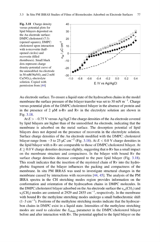
versus potential plots of the DMPC:cholesterol bilayer in the absence of protein and
in the presence of 2 μM n-Rv and Rv in the electrolyte solution are shown in
Fig. 3.18.
At E > À0.75 V versus Ag/AgCl the charge densities of the Au electrode covered
by lipid bilayers are higher than of the unmodified Au electrode, indicating that the
membrane is adsorbed on the metal surface. The desorption potential of lipid
bilayers does not depend on the presence of recoverin in the electrolyte solution.
Surface charge densities of the Au electrode modified with the DMPC: cholesterol
bilayer range from À5 to 25 μC cm
À2 (Fig. 3.18). At E < 0.0 V charge densities in
the lipid bilayer with n-Rv are comparable to those of DMPC:cholesterol bilayer. At
E ! 0.0 V charge densities decrease slightly, suggesting that n-Rv has a small impact
on the membrane structure and compactness. In the bilayer with bound Rv the
surface charge densities decrease compared to the pure lipid bilayer (Fig. 3.18).
This result indicates that the insertion of the myristoyl chain of Rv into the hydrophobic fragment of the bilayer influences the packing and compactness of the
membrane. In situ PM IRRAS was used to investigate structural changes in the
membrane caused by interactions with recoverins [44, 45]. The analysis of the PM
IRRA spectra in the CH stretching modes region provides information on the
conformation and orientation of the hydrocarbon chains in DMPC molecules. In
the DMPC:cholesterol bilayer adsorbed on the Au electrode surface the ν as (CH 2 ) and
ν s (CH 2 ) modes are centered at 2929 and 2855 cm
À1 , respectively. In the membrane
with bound Rv the methylene stretching modes undergo a small bathochromic shift
(1–3 cm
À1 ). Positions of the methylene stretching modes indicate that the hydrocarbon chains in DMPC exist in a liquid state. Intensities of the methylene stretching
modes are used to calculate the S chain parameter in the DMPC:cholesterol bilayer
before and after interaction with Rv. The potential applied to the lipid bilayer on the
-1.0
-0.8
-0.6
-0.4
-0.2
0.0
0.2
0.4
-30
-20
-10
0
10
20
30
40
M / C cm
-2
E /V vs Ag/AgCl
M
Fig. 3.18 Charge density
versus potential plots for
lipid bilayers deposited on
the Au electrode surface:
DMPC:cholesterol (7:3)
(opened squares), DMPC:
cholesterol upon interaction
with n-recoverin (halfopened circles) and
recoverin (filled
rhombuses). Small black
dots represent charge
density-potential curve of
the unmodified Au electrode
in 50 mM NaNO 3 and 2 mM
Ca(NO 3 ) 2 electrolyte
solution. Copied with
permission from [44]
3.3 In Situ PM IRRAS Studies of Films of Biomolecules Adsorbed on Electrode Surfaces 77
membrane the surface pressure of the bilayer transfer was set to 30 mN m
À1 . Charge
versus potential plots of the DMPC:cholesterol bilayer in the absence of protein and
in the presence of 2 μM n-Rv and Rv in the electrolyte solution are shown in
Fig. 3.18.
At E > À0.75 V versus Ag/AgCl the charge densities of the Au electrode covered
by lipid bilayers are higher than of the unmodified Au electrode, indicating that the
membrane is adsorbed on the metal surface. The desorption potential of lipid
bilayers does not depend on the presence of recoverin in the electrolyte solution.
Surface charge densities of the Au electrode modified with the DMPC: cholesterol
bilayer range from À5 to 25 μC cm
À2 (Fig. 3.18). At E < 0.0 V charge densities in
the lipid bilayer with n-Rv are comparable to those of DMPC:cholesterol bilayer. At
E ! 0.0 V charge densities decrease slightly, suggesting that n-Rv has a small impact
on the membrane structure and compactness. In the bilayer with bound Rv the
surface charge densities decrease compared to the pure lipid bilayer (Fig. 3.18).
This result indicates that the insertion of the myristoyl chain of Rv into the hydrophobic fragment of the bilayer influences the packing and compactness of the
membrane. In situ PM IRRAS was used to investigate structural changes in the
membrane caused by interactions with recoverins [44, 45]. The analysis of the PM
IRRA spectra in the CH stretching modes region provides information on the
conformation and orientation of the hydrocarbon chains in DMPC molecules. In
the DMPC:cholesterol bilayer adsorbed on the Au electrode surface the ν as (CH 2 ) and
ν s (CH 2 ) modes are centered at 2929 and 2855 cm
À1 , respectively. In the membrane
with bound Rv the methylene stretching modes undergo a small bathochromic shift
(1–3 cm
À1 ). Positions of the methylene stretching modes indicate that the hydrocarbon chains in DMPC exist in a liquid state. Intensities of the methylene stretching
modes are used to calculate the S chain parameter in the DMPC:cholesterol bilayer
before and after interaction with Rv. The potential applied to the lipid bilayer on the
-1.0
-0.8
-0.6
-0.4
-0.2
0.0
0.2
0.4
-30
-20
-10
0
10
20
30
40
M / C cm
-2
E /V vs Ag/AgCl
M
Fig. 3.18 Charge density
versus potential plots for
lipid bilayers deposited on
the Au electrode surface:
DMPC:cholesterol (7:3)
(opened squares), DMPC:
cholesterol upon interaction
with n-recoverin (halfopened circles) and
recoverin (filled
rhombuses). Small black
dots represent charge
density-potential curve of
the unmodified Au electrode
in 50 mM NaNO 3 and 2 mM
Ca(NO 3 ) 2 electrolyte
solution. Copied with
permission from [44]
3.3 In Situ PM IRRAS Studies of Films of Biomolecules Adsorbed on Electrode Surfaces 77