

8.7 Conclusion
The morphology and elemental composition of Cg sclerotia collected from low-pH
forest soils in central and northern Japan were analyzed by SEM-EDX. The cell wall
structure and melanin-like spherules consisted mainly of carbon, oxygen, and
aluminum. Graphite and graphene, products or by-products of substrate degradation,
were observed in the interior of sclerotia, and the graphene was adjacent to titanium
oxides or small amounts of adhering calcium, magnesium, or iron oxides. Precise
observation of the micromorphology of the interior of sclerotia grains suggests the
transformation of aluminum–carbon complexes from aluminum-rich melanin spherules to aluminum accumulated layers. This hypothesis would explain how Cg
sclerotia can resist degradation in soil and retain their structure, but it requires further
investigation.
The crystalline aluminum structures, including boehmite and aluminum sulfates,
observed in the interior of sclerotia indicate the possibility of aluminum enrichment
and mineralization. Spherical silicon particles were also observed, although their
origin (formed inside the sclerotia or contaminants) remains unclear. The activities
of microorganisms inside sclerotia could drive degradation of the sclerotia and
mineralization of their internal structure, but this too will require further
investigation.
Acknowledgements MW thanks Mr. T. Hatano, Mr. T. Hattori, and Mr. H. Terashima of JEOL
Co. Ltd., Tokyo, Japan, for technical support with SEM-EDS and micro-CT analyses.
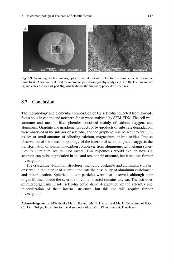
Fig. 8.9 Scanning electron micrographs of the interior of a sclerotium section, collected from the
same Iwaki A horizon soil used for micro computed tomography analysis (Fig. 8.8). The box in part
(a) indicates the area of part (b), which shows the fungal hyphae-like structures
8 Micromorphological Features of Sclerotia Grains
149
The morphology and elemental composition of Cg sclerotia collected from low-pH
forest soils in central and northern Japan were analyzed by SEM-EDX. The cell wall
structure and melanin-like spherules consisted mainly of carbon, oxygen, and
aluminum. Graphite and graphene, products or by-products of substrate degradation,
were observed in the interior of sclerotia, and the graphene was adjacent to titanium
oxides or small amounts of adhering calcium, magnesium, or iron oxides. Precise
observation of the micromorphology of the interior of sclerotia grains suggests the
transformation of aluminum–carbon complexes from aluminum-rich melanin spherules to aluminum accumulated layers. This hypothesis would explain how Cg
sclerotia can resist degradation in soil and retain their structure, but it requires further
investigation.
The crystalline aluminum structures, including boehmite and aluminum sulfates,
observed in the interior of sclerotia indicate the possibility of aluminum enrichment
and mineralization. Spherical silicon particles were also observed, although their
origin (formed inside the sclerotia or contaminants) remains unclear. The activities
of microorganisms inside sclerotia could drive degradation of the sclerotia and
mineralization of their internal structure, but this too will require further
investigation.
Acknowledgements MW thanks Mr. T. Hatano, Mr. T. Hattori, and Mr. H. Terashima of JEOL
Co. Ltd., Tokyo, Japan, for technical support with SEM-EDS and micro-CT analyses.
Fig. 8.9 Scanning electron micrographs of the interior of a sclerotium section, collected from the
same Iwaki A horizon soil used for micro computed tomography analysis (Fig. 8.8). The box in part
(a) indicates the area of part (b), which shows the fungal hyphae-like structures
8 Micromorphological Features of Sclerotia Grains
149