

8.5 Spherical Silica Particles
Spherical silica particles of 3–4 μm in diameter are frequently observed inside
sclerotia grains found from volcanic ash soils (Watanabe et al. 2004). Figure 8.7
shows the small spherical silicon particles (diameter 200 nm, O/Si ¼ 6:1) observed
in the cell wall structure of the Myoko surface soil sample (1A), and the larger
spherical particles (10 μm, O/Si ¼ 4:1) observed in the cell wall structure (C/O/
Al ¼ 68:30:2) of sclerotia collected from the Myoko buried soil horizon, 3A
(Chap. 7).
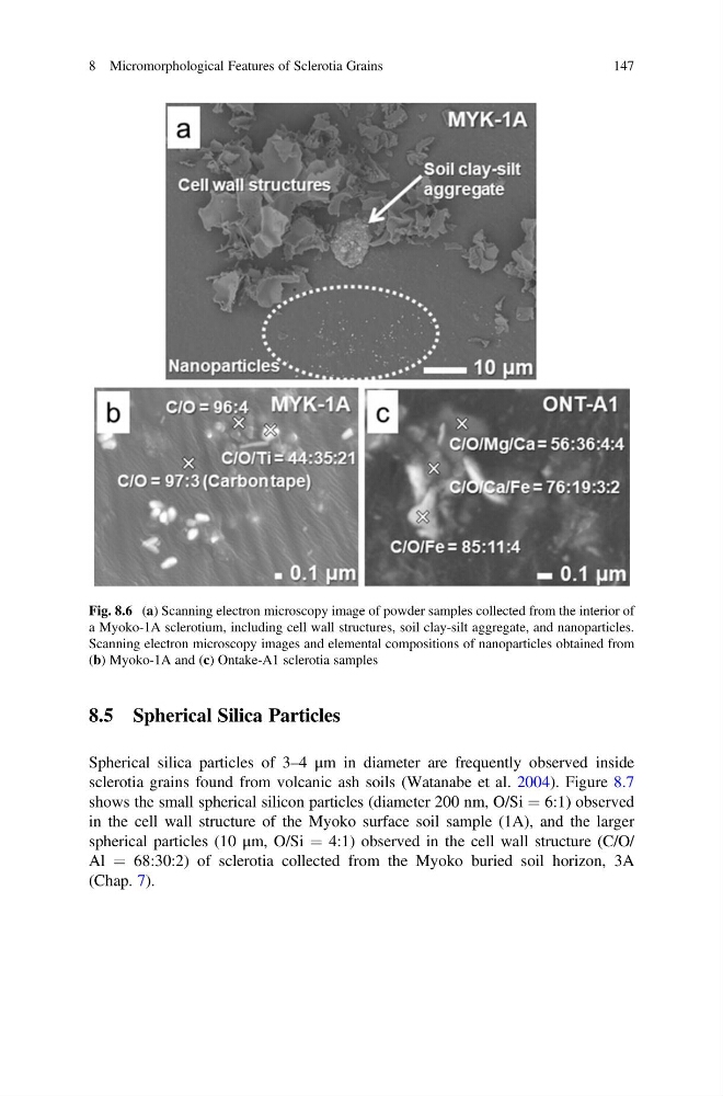
Fig. 8.6 (a) Scanning electron microscopy image of powder samples collected from the interior of
a Myoko-1A sclerotium, including cell wall structures, soil clay-silt aggregate, and nanoparticles.
Scanning electron microscopy images and elemental compositions of nanoparticles obtained from
(b) Myoko-1A and (c) Ontake-A1 sclerotia samples
8 Micromorphological Features of Sclerotia Grains
147
Spherical silica particles of 3–4 μm in diameter are frequently observed inside
sclerotia grains found from volcanic ash soils (Watanabe et al. 2004). Figure 8.7
shows the small spherical silicon particles (diameter 200 nm, O/Si ¼ 6:1) observed
in the cell wall structure of the Myoko surface soil sample (1A), and the larger
spherical particles (10 μm, O/Si ¼ 4:1) observed in the cell wall structure (C/O/
Al ¼ 68:30:2) of sclerotia collected from the Myoko buried soil horizon, 3A
(Chap. 7).
Fig. 8.6 (a) Scanning electron microscopy image of powder samples collected from the interior of
a Myoko-1A sclerotium, including cell wall structures, soil clay-silt aggregate, and nanoparticles.
Scanning electron microscopy images and elemental compositions of nanoparticles obtained from
(b) Myoko-1A and (c) Ontake-A1 sclerotia samples
8 Micromorphological Features of Sclerotia Grains
147