

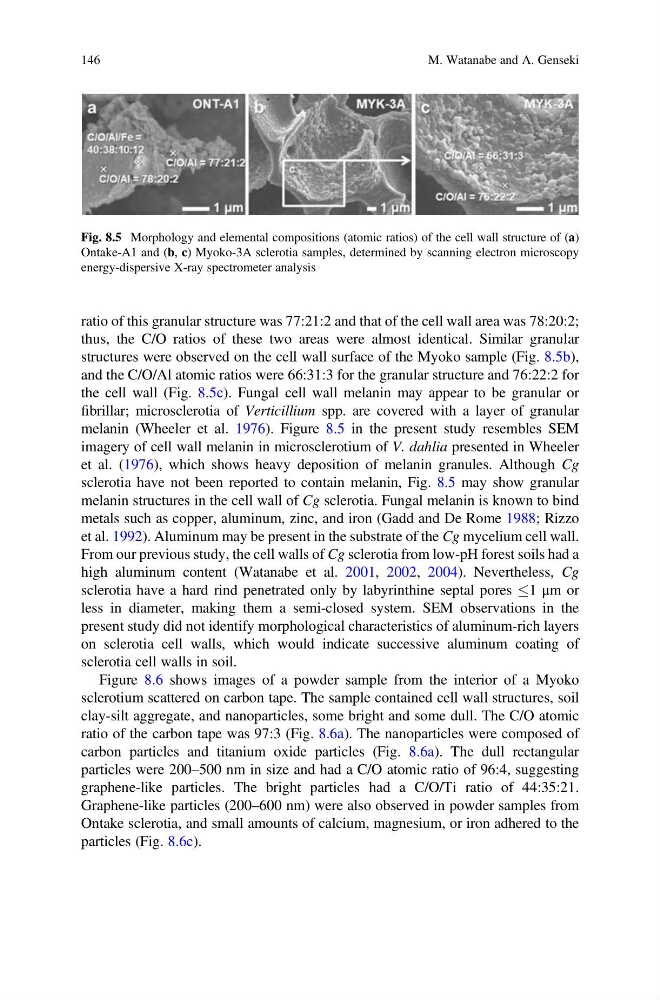
ratio of this granular structure was 77:21:2 and that of the cell wall area was 78:20:2;
thus, the C/O ratios of these two areas were almost identical. Similar granular
structures were observed on the cell wall surface of the Myoko sample (Fig. 8.5b),
and the C/O/Al atomic ratios were 66:31:3 for the granular structure and 76:22:2 for
the cell wall (Fig. 8.5c). Fungal cell wall melanin may appear to be granular or
fibrillar; microsclerotia of Verticillium spp. are covered with a layer of granular
melanin (Wheeler et al. 1976). Figure 8.5 in the present study resembles SEM
imagery of cell wall melanin in microsclerotium of V. dahlia presented in Wheeler
et al. (1976), which shows heavy deposition of melanin granules. Although Cg
sclerotia have not been reported to contain melanin, Fig. 8.5 may show granular
melanin structures in the cell wall of Cg sclerotia. Fungal melanin is known to bind
metals such as copper, aluminum, zinc, and iron (Gadd and De Rome 1988; Rizzo
et al. 1992). Aluminum may be present in the substrate of the Cg mycelium cell wall.
From our previous study, the cell walls of Cg sclerotia from low-pH forest soils had a
high aluminum content (Watanabe et al. 2001, 2002, 2004). Nevertheless, Cg
sclerotia have a hard rind penetrated only by labyrinthine septal pores 1 μm or
less in diameter, making them a semi-closed system. SEM observations in the
present study did not identify morphological characteristics of aluminum-rich layers
on sclerotia cell walls, which would indicate successive aluminum coating of
sclerotia cell walls in soil.
Figure 8.6 shows images of a powder sample from the interior of a Myoko
sclerotium scattered on carbon tape. The sample contained cell wall structures, soil
clay-silt aggregate, and nanoparticles, some bright and some dull. The C/O atomic
ratio of the carbon tape was 97:3 (Fig. 8.6a). The nanoparticles were composed of
carbon particles and titanium oxide particles (Fig. 8.6a). The dull rectangular
particles were 200–500 nm in size and had a C/O atomic ratio of 96:4, suggesting
graphene-like particles. The bright particles had a C/O/Ti ratio of 44:35:21.
Graphene-like particles (200–600 nm) were also observed in powder samples from
Ontake sclerotia, and small amounts of calcium, magnesium, or iron adhered to the
particles (Fig. 8.6c).
Fig. 8.5 Morphology and elemental compositions (atomic ratios) of the cell wall structure of (a)
Ontake-A1 and (b, c) Myoko-3A sclerotia samples, determined by scanning electron microscopy
energy-dispersive X-ray spectrometer analysis
146
M. Watanabe and A. Genseki
thus, the C/O ratios of these two areas were almost identical. Similar granular
structures were observed on the cell wall surface of the Myoko sample (Fig. 8.5b),
and the C/O/Al atomic ratios were 66:31:3 for the granular structure and 76:22:2 for
the cell wall (Fig. 8.5c). Fungal cell wall melanin may appear to be granular or
fibrillar; microsclerotia of Verticillium spp. are covered with a layer of granular
melanin (Wheeler et al. 1976). Figure 8.5 in the present study resembles SEM
imagery of cell wall melanin in microsclerotium of V. dahlia presented in Wheeler
et al. (1976), which shows heavy deposition of melanin granules. Although Cg
sclerotia have not been reported to contain melanin, Fig. 8.5 may show granular
melanin structures in the cell wall of Cg sclerotia. Fungal melanin is known to bind
metals such as copper, aluminum, zinc, and iron (Gadd and De Rome 1988; Rizzo
et al. 1992). Aluminum may be present in the substrate of the Cg mycelium cell wall.
From our previous study, the cell walls of Cg sclerotia from low-pH forest soils had a
high aluminum content (Watanabe et al. 2001, 2002, 2004). Nevertheless, Cg
sclerotia have a hard rind penetrated only by labyrinthine septal pores 1 μm or
less in diameter, making them a semi-closed system. SEM observations in the
present study did not identify morphological characteristics of aluminum-rich layers
on sclerotia cell walls, which would indicate successive aluminum coating of
sclerotia cell walls in soil.
Figure 8.6 shows images of a powder sample from the interior of a Myoko
sclerotium scattered on carbon tape. The sample contained cell wall structures, soil
clay-silt aggregate, and nanoparticles, some bright and some dull. The C/O atomic
ratio of the carbon tape was 97:3 (Fig. 8.6a). The nanoparticles were composed of
carbon particles and titanium oxide particles (Fig. 8.6a). The dull rectangular
particles were 200–500 nm in size and had a C/O atomic ratio of 96:4, suggesting
graphene-like particles. The bright particles had a C/O/Ti ratio of 44:35:21.
Graphene-like particles (200–600 nm) were also observed in powder samples from
Ontake sclerotia, and small amounts of calcium, magnesium, or iron adhered to the
particles (Fig. 8.6c).
Fig. 8.5 Morphology and elemental compositions (atomic ratios) of the cell wall structure of (a)
Ontake-A1 and (b, c) Myoko-3A sclerotia samples, determined by scanning electron microscopy
energy-dispersive X-ray spectrometer analysis
146
M. Watanabe and A. Genseki