

d3 ¼ 0.133, and d4 ¼ 0.114 for the diffraction points located on the four readable
rings. Based on the relative brightness of the points, equivalent to the relative
intensity of the X-rays (I), these four lattice distances are concordant with X-ray
powder diffraction data for boehmite, γ-AlOOH. Nevertheless, the lattice distance
expected for hkl ¼ 120 (I ¼ 65) was not confirmed here, probably due to the absence
of a specific diffraction point behind the aggregate of microcrystalline structures.
In general, boehmite consists of aluminum oxyhydroxides present in many types
of bauxites and is normally the ultimate product of intensive weathering of primary
aluminum silicates in soils (Hsu 1989). Experimental alteration of obsidian (volcanic
glass) conducted by Kawano and Tomita (1993) confirmed that boehmite has a
fibrous structure corresponding to alteration products associated with spherical
kaolinite and precursors of smectite in Al
3+ -enriched solution. On the other hand,
the number of ectomycorrhizal tips formed by Cg has been reported to increase in
soils exposed to simulated rain with a pH of 2.5 by Meira et al. (1989). Consequently, formation of Al oxyhydroxide polymorphs in sclerotia grains might result
from Al dissolution–precipitation inside the grains. A mycelium-like structure was
observed in the central part of the sclerotia grains, and a biochemical process
performed by fungi colonizing the sclerotia might be involved in the dissolution of
aluminum from the matrix of the grain, in the form of aluminum–carbon complexes.
This might induce aluminum saturation and precipitation under acidic conditions,
which could lead to formation of boehmite-like aluminum oxyhydroxides.
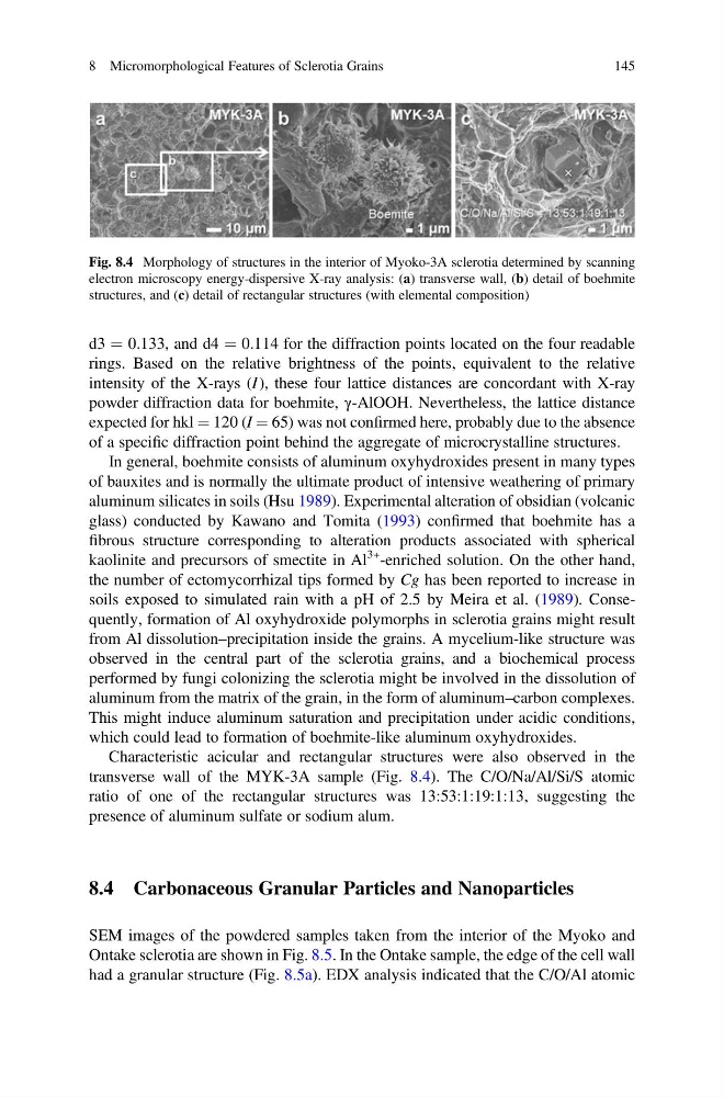
Characteristic acicular and rectangular structures were also observed in the
transverse wall of the MYK-3A sample (Fig. 8.4). The C/O/Na/Al/Si/S atomic
ratio of one of the rectangular structures was 13:53:1:19:1:13, suggesting the
presence of aluminum sulfate or sodium alum.
8.4 Carbonaceous Granular Particles and Nanoparticles
SEM images of the powdered samples taken from the interior of the Myoko and
Ontake sclerotia are shown in Fig. 8.5. In the Ontake sample, the edge of the cell wall
had a granular structure (Fig. 8.5a). EDX analysis indicated that the C/O/Al atomic
Fig. 8.4 Morphology of structures in the interior of Myoko-3A sclerotia determined by scanning
electron microscopy energy-dispersive X-ray analysis: (a) transverse wall, (b) detail of boehmite
structures, and (c) detail of rectangular structures (with elemental composition)
8 Micromorphological Features of Sclerotia Grains
145
rings. Based on the relative brightness of the points, equivalent to the relative
intensity of the X-rays (I), these four lattice distances are concordant with X-ray
powder diffraction data for boehmite, γ-AlOOH. Nevertheless, the lattice distance
expected for hkl ¼ 120 (I ¼ 65) was not confirmed here, probably due to the absence
of a specific diffraction point behind the aggregate of microcrystalline structures.
In general, boehmite consists of aluminum oxyhydroxides present in many types
of bauxites and is normally the ultimate product of intensive weathering of primary
aluminum silicates in soils (Hsu 1989). Experimental alteration of obsidian (volcanic
glass) conducted by Kawano and Tomita (1993) confirmed that boehmite has a
fibrous structure corresponding to alteration products associated with spherical
kaolinite and precursors of smectite in Al
3+ -enriched solution. On the other hand,
the number of ectomycorrhizal tips formed by Cg has been reported to increase in
soils exposed to simulated rain with a pH of 2.5 by Meira et al. (1989). Consequently, formation of Al oxyhydroxide polymorphs in sclerotia grains might result
from Al dissolution–precipitation inside the grains. A mycelium-like structure was
observed in the central part of the sclerotia grains, and a biochemical process
performed by fungi colonizing the sclerotia might be involved in the dissolution of
aluminum from the matrix of the grain, in the form of aluminum–carbon complexes.
This might induce aluminum saturation and precipitation under acidic conditions,
which could lead to formation of boehmite-like aluminum oxyhydroxides.
Characteristic acicular and rectangular structures were also observed in the
transverse wall of the MYK-3A sample (Fig. 8.4). The C/O/Na/Al/Si/S atomic
ratio of one of the rectangular structures was 13:53:1:19:1:13, suggesting the
presence of aluminum sulfate or sodium alum.
8.4 Carbonaceous Granular Particles and Nanoparticles
SEM images of the powdered samples taken from the interior of the Myoko and
Ontake sclerotia are shown in Fig. 8.5. In the Ontake sample, the edge of the cell wall
had a granular structure (Fig. 8.5a). EDX analysis indicated that the C/O/Al atomic
Fig. 8.4 Morphology of structures in the interior of Myoko-3A sclerotia determined by scanning
electron microscopy energy-dispersive X-ray analysis: (a) transverse wall, (b) detail of boehmite
structures, and (c) detail of rectangular structures (with elemental composition)
8 Micromorphological Features of Sclerotia Grains
145