

the transverse wall associated with a septal pore (Fig. 8.2g). At a higher magnification, the micromorphology of the matrix had a honeycomb structure with a chemical
composition of 2:1 O:Al (Fig. 8.2h, i). Metal elements such as nickel and lead were
frequently detected as roughly spherical structures intermingled with the matrix
(Fig. 8.2i).
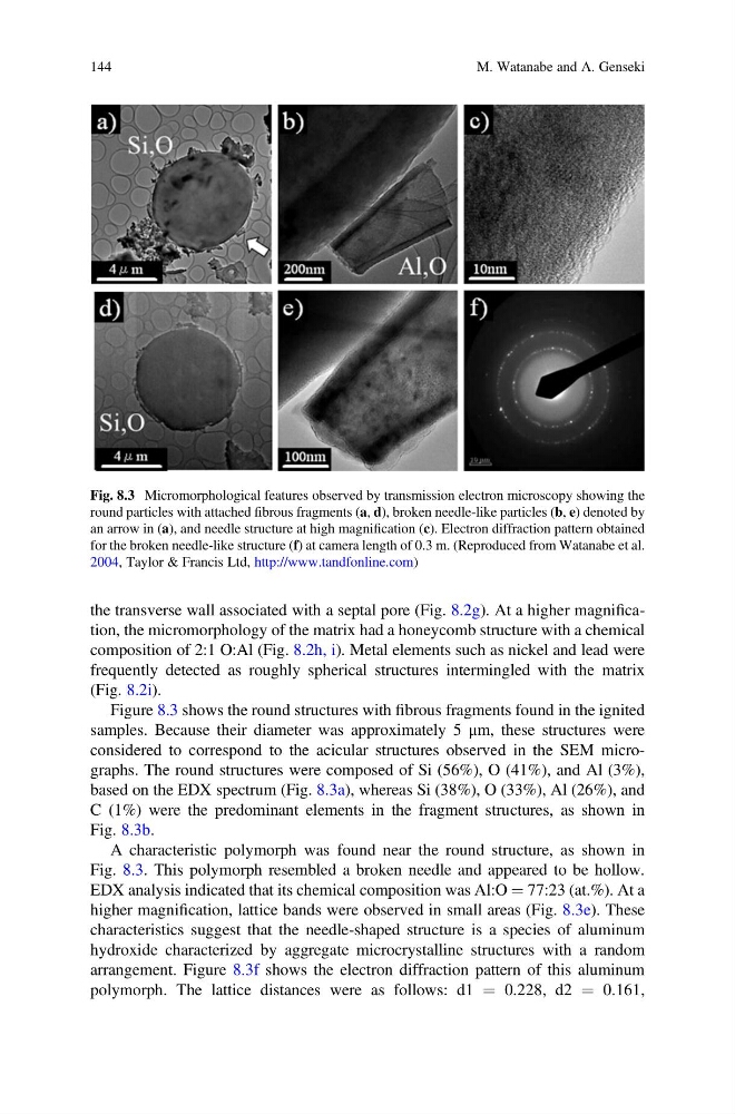
Figure 8.3 shows the round structures with fibrous fragments found in the ignited
samples. Because their diameter was approximately 5 μm, these structures were
considered to correspond to the acicular structures observed in the SEM micrographs. The round structures were composed of Si (56%), O (41%), and Al (3%),
based on the EDX spectrum (Fig. 8.3a), whereas Si (38%), O (33%), Al (26%), and
C (1%) were the predominant elements in the fragment structures, as shown in
Fig. 8.3b.
A characteristic polymorph was found near the round structure, as shown in
Fig. 8.3. This polymorph resembled a broken needle and appeared to be hollow.
EDX analysis indicated that its chemical composition was Al:O ¼ 77:23 (at.%). At a
higher magnification, lattice bands were observed in small areas (Fig. 8.3e). These
characteristics suggest that the needle-shaped structure is a species of aluminum
hydroxide characterized by aggregate microcrystalline structures with a random
arrangement. Figure 8.3f shows the electron diffraction pattern of this aluminum
polymorph. The lattice distances were as follows: d1 ¼ 0.228, d2 ¼ 0.161,
Fig. 8.3 Micromorphological features observed by transmission electron microscopy showing the
round particles with attached fibrous fragments (a, d), broken needle-like particles (b, e) denoted by
an arrow in (a), and needle structure at high magnification (c). Electron diffraction pattern obtained
for the broken needle-like structure (f) at camera length of 0.3 m. (Reproduced from Watanabe et al.
2004, Taylor & Francis Ltd, http://www.tandfonline.com)
144
M. Watanabe and A. Genseki
composition of 2:1 O:Al (Fig. 8.2h, i). Metal elements such as nickel and lead were
frequently detected as roughly spherical structures intermingled with the matrix
(Fig. 8.2i).
Figure 8.3 shows the round structures with fibrous fragments found in the ignited
samples. Because their diameter was approximately 5 μm, these structures were
considered to correspond to the acicular structures observed in the SEM micrographs. The round structures were composed of Si (56%), O (41%), and Al (3%),
based on the EDX spectrum (Fig. 8.3a), whereas Si (38%), O (33%), Al (26%), and
C (1%) were the predominant elements in the fragment structures, as shown in
Fig. 8.3b.
A characteristic polymorph was found near the round structure, as shown in
Fig. 8.3. This polymorph resembled a broken needle and appeared to be hollow.
EDX analysis indicated that its chemical composition was Al:O ¼ 77:23 (at.%). At a
higher magnification, lattice bands were observed in small areas (Fig. 8.3e). These
characteristics suggest that the needle-shaped structure is a species of aluminum
hydroxide characterized by aggregate microcrystalline structures with a random
arrangement. Figure 8.3f shows the electron diffraction pattern of this aluminum
polymorph. The lattice distances were as follows: d1 ¼ 0.228, d2 ¼ 0.161,
Fig. 8.3 Micromorphological features observed by transmission electron microscopy showing the
round particles with attached fibrous fragments (a, d), broken needle-like particles (b, e) denoted by
an arrow in (a), and needle structure at high magnification (c). Electron diffraction pattern obtained
for the broken needle-like structure (f) at camera length of 0.3 m. (Reproduced from Watanabe et al.
2004, Taylor & Francis Ltd, http://www.tandfonline.com)
144
M. Watanabe and A. Genseki