

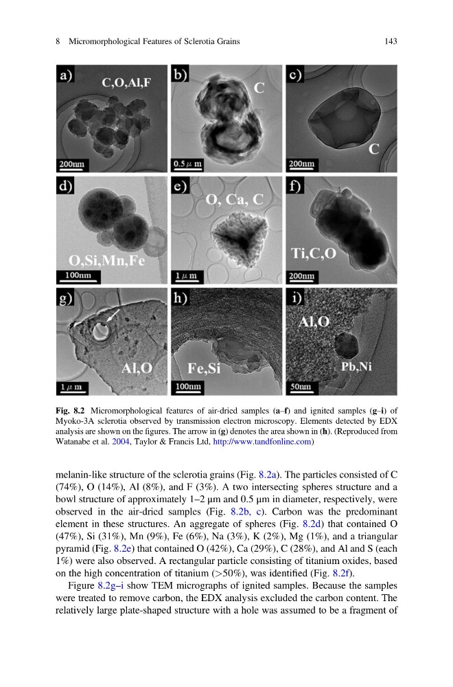
melanin-like structure of the sclerotia grains (Fig. 8.2a). The particles consisted of C
(74%), O (14%), Al (8%), and F (3%). A two intersecting spheres structure and a
bowl structure of approximately 1–2 μm and 0.5 μm in diameter, respectively, were
observed in the air-dried samples (Fig. 8.2b, c). Carbon was the predominant
element in these structures. An aggregate of spheres (Fig. 8.2d) that contained O
(47%), Si (31%), Mn (9%), Fe (6%), Na (3%), K (2%), Mg (1%), and a triangular
pyramid (Fig. 8.2e) that contained O (42%), Ca (29%), C (28%), and Al and S (each
1%) were also observed. A rectangular particle consisting of titanium oxides, based
on the high concentration of titanium (>50%), was identified (Fig. 8.2f).
Figure 8.2g–i show TEM micrographs of ignited samples. Because the samples
were treated to remove carbon, the EDX analysis excluded the carbon content. The
relatively large plate-shaped structure with a hole was assumed to be a fragment of
Fig. 8.2 Micromorphological features of air-dried samples (a–f) and ignited samples (g–i) of
Myoko-3A sclerotia observed by transmission electron microscopy. Elements detected by EDX
analysis are shown on the figures. The arrow in (g) denotes the area shown in (h). (Reproduced from
Watanabe et al. 2004, Taylor & Francis Ltd, http://www.tandfonline.com)
8 Micromorphological Features of Sclerotia Grains
143
(74%), O (14%), Al (8%), and F (3%). A two intersecting spheres structure and a
bowl structure of approximately 1–2 μm and 0.5 μm in diameter, respectively, were
observed in the air-dried samples (Fig. 8.2b, c). Carbon was the predominant
element in these structures. An aggregate of spheres (Fig. 8.2d) that contained O
(47%), Si (31%), Mn (9%), Fe (6%), Na (3%), K (2%), Mg (1%), and a triangular
pyramid (Fig. 8.2e) that contained O (42%), Ca (29%), C (28%), and Al and S (each
1%) were also observed. A rectangular particle consisting of titanium oxides, based
on the high concentration of titanium (>50%), was identified (Fig. 8.2f).
Figure 8.2g–i show TEM micrographs of ignited samples. Because the samples
were treated to remove carbon, the EDX analysis excluded the carbon content. The
relatively large plate-shaped structure with a hole was assumed to be a fragment of
Fig. 8.2 Micromorphological features of air-dried samples (a–f) and ignited samples (g–i) of
Myoko-3A sclerotia observed by transmission electron microscopy. Elements detected by EDX
analysis are shown on the figures. The arrow in (g) denotes the area shown in (h). (Reproduced from
Watanabe et al. 2004, Taylor & Francis Ltd, http://www.tandfonline.com)
8 Micromorphological Features of Sclerotia Grains
143