

A single sclerotium grain from Iwaki A horizon soil was used for micro computed
tomography (micro-CT) analysis (μCT50, Scanco Medical AG, Switzerland) to
observe the inner structure in a non-destructive manner. Image processing software
(Scanco Medical AG IPL) was used to obtain three-dimensional images. The
measurement conditions were as follows: X-ray tube voltage/current: 45 kV/
88 μA; air, no filter; pixel resolution: (8192 Â 8192); slice thickness: 0.5 μm;
exposure time: 1500 ms; sample rotation angle: 0.12
; integration number:
10 times; total number of images: 2188.
8.3 Aluminum Hydroxide Polymorphs
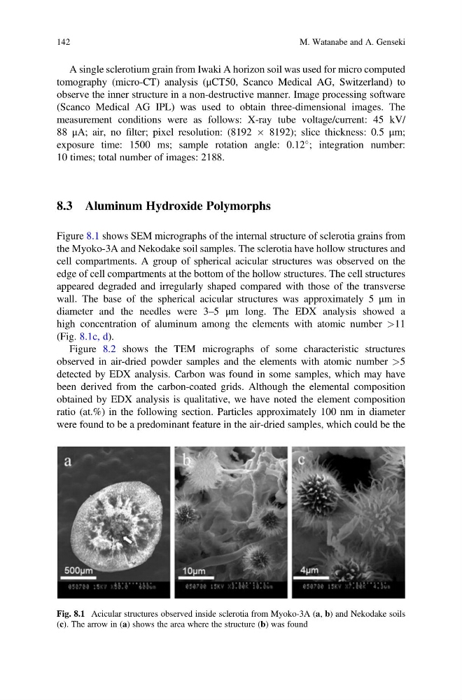
Figure 8.1 shows SEM micrographs of the internal structure of sclerotia grains from
the Myoko-3A and Nekodake soil samples. The sclerotia have hollow structures and
cell compartments. A group of spherical acicular structures was observed on the
edge of cell compartments at the bottom of the hollow structures. The cell structures
appeared degraded and irregularly shaped compared with those of the transverse
wall. The base of the spherical acicular structures was approximately 5 μm in
diameter and the needles were 3–5 μm long. The EDX analysis showed a
high concentration of aluminum among the elements with atomic number >11
(Fig. 8.1c, d).
Figure 8.2 shows the TEM micrographs of some characteristic structures
observed in air-dried powder samples and the elements with atomic number >5
detected by EDX analysis. Carbon was found in some samples, which may have
been derived from the carbon-coated grids. Although the elemental composition
obtained by EDX analysis is qualitative, we have noted the element composition
ratio (at.%) in the following section. Particles approximately 100 nm in diameter
were found to be a predominant feature in the air-dried samples, which could be the
Fig. 8.1 Acicular structures observed inside sclerotia from Myoko-3A (a, b) and Nekodake soils
(c). The arrow in (a) shows the area where the structure (b) was found
142
M. Watanabe and A. Genseki
tomography (micro-CT) analysis (μCT50, Scanco Medical AG, Switzerland) to
observe the inner structure in a non-destructive manner. Image processing software
(Scanco Medical AG IPL) was used to obtain three-dimensional images. The
measurement conditions were as follows: X-ray tube voltage/current: 45 kV/
88 μA; air, no filter; pixel resolution: (8192 Â 8192); slice thickness: 0.5 μm;
exposure time: 1500 ms; sample rotation angle: 0.12
; integration number:
10 times; total number of images: 2188.
8.3 Aluminum Hydroxide Polymorphs
Figure 8.1 shows SEM micrographs of the internal structure of sclerotia grains from
the Myoko-3A and Nekodake soil samples. The sclerotia have hollow structures and
cell compartments. A group of spherical acicular structures was observed on the
edge of cell compartments at the bottom of the hollow structures. The cell structures
appeared degraded and irregularly shaped compared with those of the transverse
wall. The base of the spherical acicular structures was approximately 5 μm in
diameter and the needles were 3–5 μm long. The EDX analysis showed a
high concentration of aluminum among the elements with atomic number >11
(Fig. 8.1c, d).
Figure 8.2 shows the TEM micrographs of some characteristic structures
observed in air-dried powder samples and the elements with atomic number >5
detected by EDX analysis. Carbon was found in some samples, which may have
been derived from the carbon-coated grids. Although the elemental composition
obtained by EDX analysis is qualitative, we have noted the element composition
ratio (at.%) in the following section. Particles approximately 100 nm in diameter
were found to be a predominant feature in the air-dried samples, which could be the
Fig. 8.1 Acicular structures observed inside sclerotia from Myoko-3A (a, b) and Nekodake soils
(c). The arrow in (a) shows the area where the structure (b) was found
142
M. Watanabe and A. Genseki