

134 unifying physics of accelerators, lasers and plasma
which correspond to the maximal reflection of X-rays from
the crystal. In the above equation, n is an integer, d is the
distance between the crystal planes and θ is the angle between X-rays and scattering crystal planes. The monochromator plates are usually made from crystals of Si or Ge. The
symmetrical configuration shown in Fig. 7.7.a is standard,
while the asymmetrical one (Fig. 7.7.b) allows for the increase
in angular resolution — thus narrowing the resulting energy
spread of the X-ray beam.
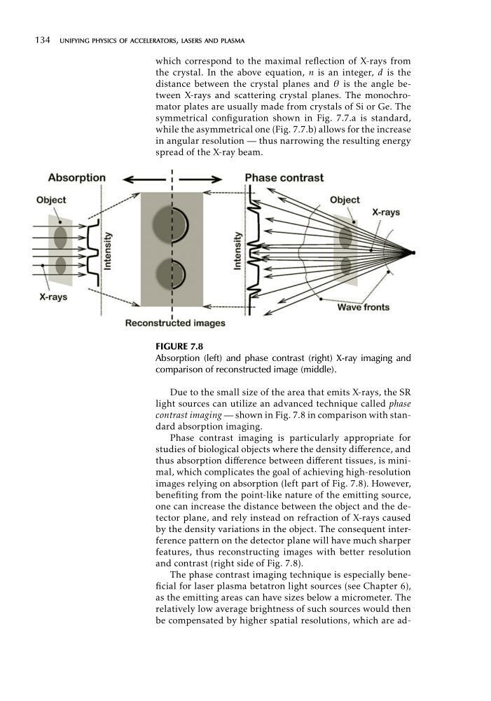
FIGURE 7.8
Absorption (left) and phase contrast (right) X-ray imaging and
comparison of reconstructed image (middle).
Due to the small size of the area that emits X-rays, the SR
light sources can utilize an advanced technique called phase
contrast imaging — shown in Fig. 7.8 in comparison with standard absorption imaging.
Phase contrast imaging is particularly appropriate for
studies of biological objects where the density difference, and
thus absorption difference between different tissues, is minimal, which complicates the goal of achieving high-resolution
images relying on absorption (left part of Fig. 7.8). However,
benefiting from the point-like nature of the emitting source,
one can increase the distance between the object and the detector plane, and rely instead on refraction of X-rays caused
by the density variations in the object. The consequent interference pattern on the detector plane will have much sharper
features, thus reconstructing images with better resolution
and contrast (right side of Fig. 7.8).
The phase contrast imaging technique is especially beneficial for laser plasma betatron light sources (see Chapter 6),
as the emitting areas can have sizes below a micrometer. The
relatively low average brightness of such sources would then
be compensated by higher spatial resolutions, which are ad
which correspond to the maximal reflection of X-rays from
the crystal. In the above equation, n is an integer, d is the
distance between the crystal planes and θ is the angle between X-rays and scattering crystal planes. The monochromator plates are usually made from crystals of Si or Ge. The
symmetrical configuration shown in Fig. 7.7.a is standard,
while the asymmetrical one (Fig. 7.7.b) allows for the increase
in angular resolution — thus narrowing the resulting energy
spread of the X-ray beam.
FIGURE 7.8
Absorption (left) and phase contrast (right) X-ray imaging and
comparison of reconstructed image (middle).
Due to the small size of the area that emits X-rays, the SR
light sources can utilize an advanced technique called phase
contrast imaging — shown in Fig. 7.8 in comparison with standard absorption imaging.
Phase contrast imaging is particularly appropriate for
studies of biological objects where the density difference, and
thus absorption difference between different tissues, is minimal, which complicates the goal of achieving high-resolution
images relying on absorption (left part of Fig. 7.8). However,
benefiting from the point-like nature of the emitting source,
one can increase the distance between the object and the detector plane, and rely instead on refraction of X-rays caused
by the density variations in the object. The consequent interference pattern on the detector plane will have much sharper
features, thus reconstructing images with better resolution
and contrast (right side of Fig. 7.8).
The phase contrast imaging technique is especially beneficial for laser plasma betatron light sources (see Chapter 6),
as the emitting areas can have sizes below a micrometer. The
relatively low average brightness of such sources would then
be compensated by higher spatial resolutions, which are ad