

pulse strength (V/m)
500
400
300
200
100
t 1
= 1 μ
s e c
t 1
= 5 0 μ
s e c
0
50
100
Incident pulse strength (kV/m)
Peak transmitted (in muscle)
48
Electromagnetic Fields in Biological Systems
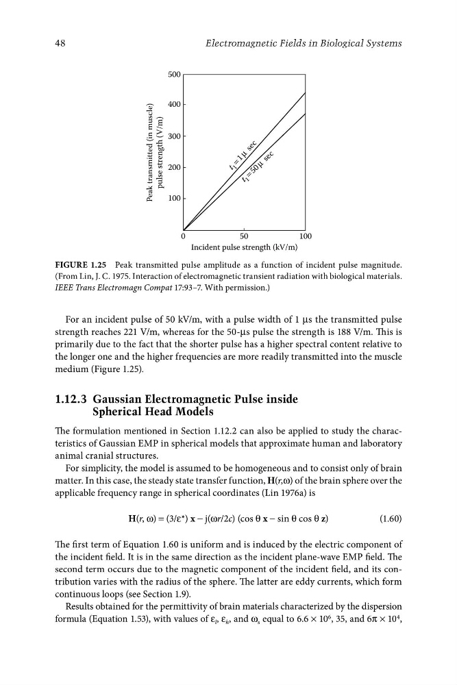
FigurE 1.25 Peak transmitted pulse amplitude as a function of incident pulse magnitude.
(From Lin, J. C. 1975. Interaction of electromagnetic transient radiation with biological materials.
IEEE Trans Electromagn Compat 17:93–7. With permission.)
For an incident pulse of 50 kV/m, with a pulse width of 1 μs the transmitted pulse
strength reaches 221 V/m, whereas for the 50-μs pulse the strength is 188 V/m. This is
primarily due to the fact that the shorter pulse has a higher spectral content relative to
the longer one and the higher frequencies are more readily transmitted into the muscle
medium (Figure 1.25).
1.12.3 Gaussian Electromagnetic Pulse inside
Spherical Head Models
The formulation mentioned in Section 1.12.2 can also be applied to study the characteristics of Gaussian EMP in spherical models that approximate human and laboratory
animal cranial structures.
For simplicity, the model is assumed to be homogeneous and to consist only of brain
matter. In this case, the steady state transfer function, H(r,ω) of the brain sphere over the
applicable frequency range in spherical coordinates (Lin 1976a) is
H(r, ω) = (3/ε*) x − j(ωr/2c) (cos θ x − sin θ cos θ z)
(1.60)
The first term of Equation 1.60 is uniform and is induced by the electric component of
the incident field. It is in the same direction as the incident plane-wave EMP field. The
second term occurs due to the magnetic component of the incident field, and its contribution varies with the radius of the sphere. The latter are eddy currents, which form
continuous loops (see Section 1.9).
Results obtained for the permittivity of brain materials characterized by the dispersion
formula (Equation 1.53), with values of ε l , ε h , and ω s equal to 6.6 × 10 6 , 35, and 6π × 10 4 ,