

Transmitted pulse (V/m) (×10
3
)
.03
.01
.05
.07
−.01
−.03
−.10
−.08
−.02
.02
.08
.10
× 4
R = 10 cm
T 1 = 1 microsec
R = 5 cm
R = 0 cm
R = −5 cm
R = −10 cm
Time (sec) ( 10 )
T 1 = 1 microsec
−.03
−.05
−.03
−.01
.01
.03
.05
R = 3.5 cm
R = 1.75 cm
R = 0 cm
R = −1.75 cm
R = −3.5 cm
Transmitted pulse (V/m) (×10 3
)
−.01
.01
.03
.05
.07
Time (sec) (×10
4 )
49
Coupling of Electromagnetic Fields into Biological Systems
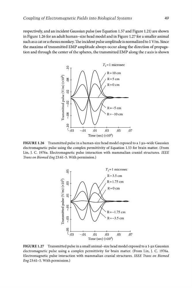
respectively, and an incident Gaussian pulse (see Equation 1.57 and Figure 1.21) are shown
in Figure 1.26 for an adult human−size head model and in Figure 1.27 for a smaller animal
such as a cat or a rhesus monkey. The incident pulse amplitude is normalized to 1 V/m. Since
the maxima of transmitted EMP amplitude always occur along the direction of propagation and through the center of the spheres, the transmitted EMP along the z axis is shown
FigurE 1.26 Transmitted pulse in a human-size head model exposed to a 1 μs–wide Gaussian
electromagnetic pulse using the complex permittivity of Equation 1.53 for brain matter. (From
Lin, J. C. 1976a. Electromagnetic pulse interaction with mammalian craniel structures. IEEE
Trans on Biomed Eng 23:61–5. With permission.)
FigurE 1.27 Transmitted pulse in a small animal–size head model exposed to a 1-μs Gaussian
electromagnetic pulse using a complex permittivity for brain matter. (From Lin, J. C. 1976a.
Electromagnetic pulse interaction with mammalian cranial structures. IEEE Trans on Biomed
Eng 23:61–5. With permission.)