

391
Terahertz Radiation
visualization of this banding pattern, such as polarization and transmission electron
microscopy (TEM), are commonly used for tissue damage evaluation (See Section 7.4.7
for further details regarding the practical significance of these measurements.)
7.4.2 Fundamental Principles of Terahertz-Tissue Optics
When THz photons interact with skin, a fraction of the photons are reflected at the material boundary, and the remaining photons are transmitted into the skin. Figure 7.14b is
a graphical representation of an incident THz wave being reflected and transmitted into
human skin. Assuming an unit incident irradiance E 0 (Wm −2 ), the light transmitted
into skin can be defined as: T = 1 − R, where T and R represent the ratio of transmitted
and specular reflected photons, respectively. Surface losses are primarily due to the differences in the index of refraction (n) between air and tissue, whereas the transmission
losses are governed by the tissue’s optical properties (i.e., absorption and scattering coefficients, μ a and μ s , respectively).
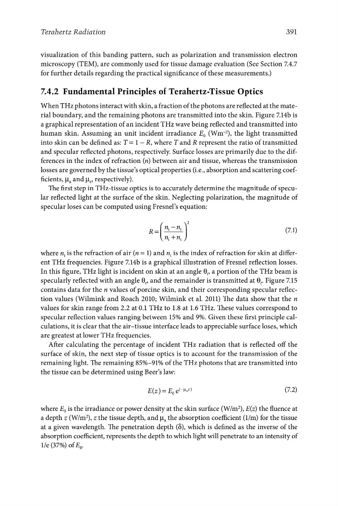
The first step in THz-tissue optics is to accurately determine the magnitude of specular reflected light at the surface of the skin. Neglecting polarization, the magnitude of
specular loses can be computed using Fresnel’s equation:
2
⎞
⎠
⎛
⎝
i
t
⎜
⎟
where n i is the refraction of air (n = 1) and n t is the index of refraction for skin at different THz frequencies. Figure 7.14b is a graphical illustration of Fresnel reflection losses.
In this figure, THz light is incident on skin at an angle θ i , a portion of the THz beam is
specularly reflected with an angle θ r , and the remainder is transmitted at θ t . Figure 7.15
contains data for the n values of porcine skin, and their corresponding specular reflection values (Wilmink and Roach 2010; Wilmink et al. 2011) The data show that the n
values for skin range from 2.2 at 0.1 THz to 1.8 at 1.6 THz. These values correspond to
specular reflection values ranging between 15% and 9%. Given these first principle calculations, it is clear that the air–tissue interface leads to appreciable surface loses, which
are greatest at lower THz frequencies.
After calculating the percentage of incident THz radiation that is reflected off the
surface of skin, the next step of tissue optics is to account for the transmission of the
remaining light. The remaining 85%–91% of the THz photons that are transmitted into
the tissue can be determined using Beer’s law:
−µ a z )
E z
( ) = E 0 e
(
(7.2)
where E 0 is the irradiance or power density at the skin surface (W/m 2 ), E(z) the fluence at
a depth z (W/m 2 ), z the tissue depth, and μ a the absorption coefficient (1/m) for the tissue
at a given wavelength. The penetration depth (δ), which is defined as the inverse of the
absorption coefficient, represents the depth to which light will penetrate to an intensity of
1/e (37%) of E 0 .
−
+
n
n i
n
n
(7.1)
R =
t