

390
Electromagnetic Fields in Biological Systems
epi
bm
d
sc
sl
sg
ss
sb
su
Subcutaneous
tissue
Artery
Vein
Lymph
vessel
n i = air
n t = tissue
θ i
θ r
θ t
Dermis
Epidermis
(a)
(b)
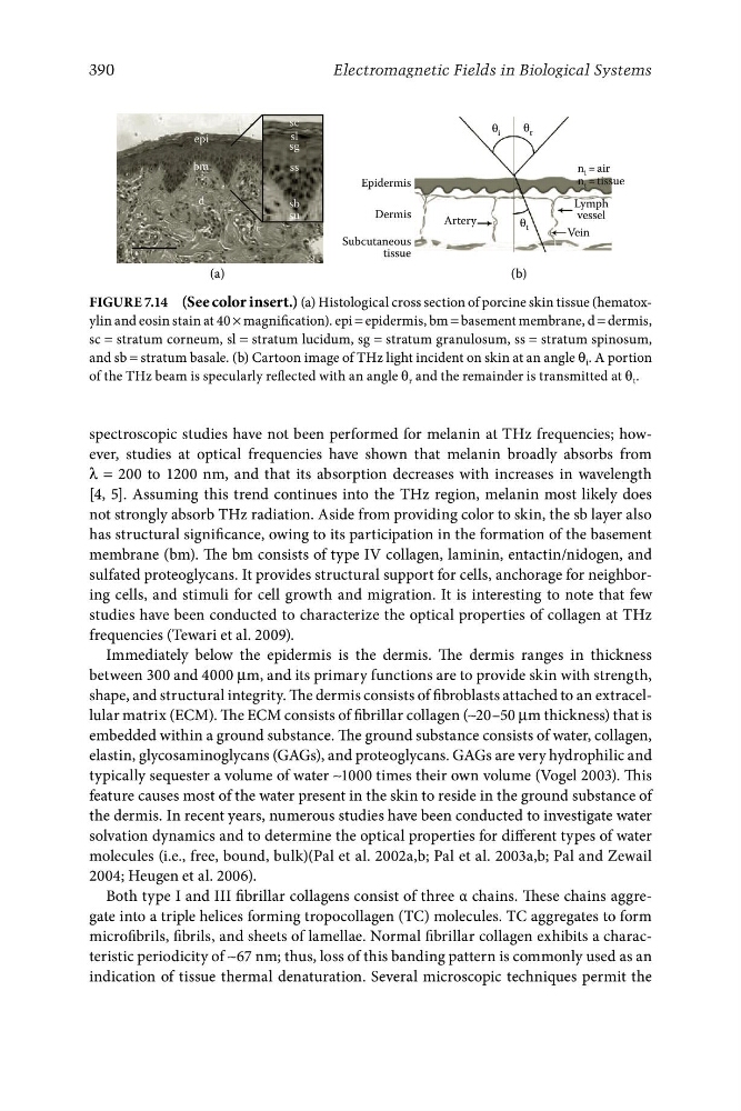
Figure 7.14 (See color insert.) (a) Histological cross section of porcine skin tissue (hematoxylin and eosin stain at 40 × magnification). epi = epidermis, bm = basement membrane, d = dermis,
sc = stratum corneum, sl = stratum lucidum, sg = stratum granulosum, ss = stratum spinosum,
and sb = stratum basale. (b) Cartoon image of THz light incident on skin at an angle θ i . A portion
of the THz beam is specularly reflected with an angle θ r and the remainder is transmitted at θ t .
spectroscopic studies have not been performed for melanin at THz frequencies; however, studies at optical frequencies have shown that melanin broadly absorbs from
λ = 200 to 1200 nm, and that its absorption decreases with increases in wavelength
[4, 5]. Assuming this trend continues into the THz region, melanin most likely does
not strongly absorb THz radiation. Aside from providing color to skin, the sb layer also
has structural significance, owing to its participation in the formation of the basement
membrane (bm). The bm consists of type IV collagen, laminin, entactin/nidogen, and
sulfated proteoglycans. It provides structural support for cells, anchorage for neighboring cells, and stimuli for cell growth and migration. It is interesting to note that few
studies have been conducted to characterize the optical properties of collagen at THz
frequencies (Tewari et al. 2009).
Immediately below the epidermis is the dermis. The dermis ranges in thickness
between 300 and 4000 μm, and its primary functions are to provide skin with strength,
shape, and structural integrity. The dermis consists of fibroblasts attached to an extracellular matrix (ECM). The ECM consists of fibrillar collagen (∼20–50 μm thickness) that is
embedded within a ground substance. The ground substance consists of water, collagen,
elastin, glycosaminoglycans (GAGs), and proteoglycans. GAGs are very hydrophilic and
typically sequester a volume of water ∼1000 times their own volume (Vogel 2003). This
feature causes most of the water present in the skin to reside in the ground substance of
the dermis. In recent years, numerous studies have been conducted to investigate water
solvation dynamics and to determine the optical properties for different types of water
molecules (i.e., free, bound, bulk)(Pal et al. 2002a,b; Pal et al. 2003a,b; Pal and Zewail
2004; Heugen et al. 2006).
Both type I and III fibrillar collagens consist of three α chains. These chains aggregate into a triple helices forming tropocollagen (TC) molecules. TC aggregates to form
microfibrils, fibrils, and sheets of lamellae. Normal fibrillar collagen exhibits a characteristic periodicity of ∼67 nm; thus, loss of this banding pattern is commonly used as an
indication of tissue thermal denaturation. Several microscopic techniques permit the
Electromagnetic Fields in Biological Systems
epi
bm
d
sc
sl
sg
ss
sb
su
Subcutaneous
tissue
Artery
Vein
Lymph
vessel
n i = air
n t = tissue
θ i
θ r
θ t
Dermis
Epidermis
(a)
(b)
Figure 7.14 (See color insert.) (a) Histological cross section of porcine skin tissue (hematoxylin and eosin stain at 40 × magnification). epi = epidermis, bm = basement membrane, d = dermis,
sc = stratum corneum, sl = stratum lucidum, sg = stratum granulosum, ss = stratum spinosum,
and sb = stratum basale. (b) Cartoon image of THz light incident on skin at an angle θ i . A portion
of the THz beam is specularly reflected with an angle θ r and the remainder is transmitted at θ t .
spectroscopic studies have not been performed for melanin at THz frequencies; however, studies at optical frequencies have shown that melanin broadly absorbs from
λ = 200 to 1200 nm, and that its absorption decreases with increases in wavelength
[4, 5]. Assuming this trend continues into the THz region, melanin most likely does
not strongly absorb THz radiation. Aside from providing color to skin, the sb layer also
has structural significance, owing to its participation in the formation of the basement
membrane (bm). The bm consists of type IV collagen, laminin, entactin/nidogen, and
sulfated proteoglycans. It provides structural support for cells, anchorage for neighboring cells, and stimuli for cell growth and migration. It is interesting to note that few
studies have been conducted to characterize the optical properties of collagen at THz
frequencies (Tewari et al. 2009).
Immediately below the epidermis is the dermis. The dermis ranges in thickness
between 300 and 4000 μm, and its primary functions are to provide skin with strength,
shape, and structural integrity. The dermis consists of fibroblasts attached to an extracellular matrix (ECM). The ECM consists of fibrillar collagen (∼20–50 μm thickness) that is
embedded within a ground substance. The ground substance consists of water, collagen,
elastin, glycosaminoglycans (GAGs), and proteoglycans. GAGs are very hydrophilic and
typically sequester a volume of water ∼1000 times their own volume (Vogel 2003). This
feature causes most of the water present in the skin to reside in the ground substance of
the dermis. In recent years, numerous studies have been conducted to investigate water
solvation dynamics and to determine the optical properties for different types of water
molecules (i.e., free, bound, bulk)(Pal et al. 2002a,b; Pal et al. 2003a,b; Pal and Zewail
2004; Heugen et al. 2006).
Both type I and III fibrillar collagens consist of three α chains. These chains aggregate into a triple helices forming tropocollagen (TC) molecules. TC aggregates to form
microfibrils, fibrils, and sheets of lamellae. Normal fibrillar collagen exhibits a characteristic periodicity of ∼67 nm; thus, loss of this banding pattern is commonly used as an
indication of tissue thermal denaturation. Several microscopic techniques permit the