

1895: The first X-ray image
1995: The first T-ray image
Freshly cut leaf
After 48 hours
Water concentration
(a)
(b)
387
Terahertz Radiation
These systems are presently being used to detect and diagnose cancer, burns, and other
disorders (Chan et al. 2007; Oh et al. 2008; Ashworth et al. 2009; Oh et al. 2009). The
contrast and sensitivity of these systems are due to the fact that the optical properties of
healthy, burned, and diseased tissues vary widely at THz frequencies. In addition, these
properties have been shown to vary with several other factors: organism (e.g., human,
rat, and porcine) (Pickwell et al. 2004; Pickwell et al. 2005; Huang et al. 2009; PickwellMacPherson and Wallace 2009; Sun et al. 2009), tissue type (e.g., skin, muscle, adipose,
liver, and heart) (Fitzgerald et al. 2003; Pickwell et al. 2004; He 2006), preparation technique (e.g., freshly excised vs. frozen–thawed), and fixation duration (Sun et al. 2009).
Overall, THz spectrometers that permit reflection-based measurements have proven to
be useful for a host of medical and biomedical applications.
7.3.2 Terahertz Imaging
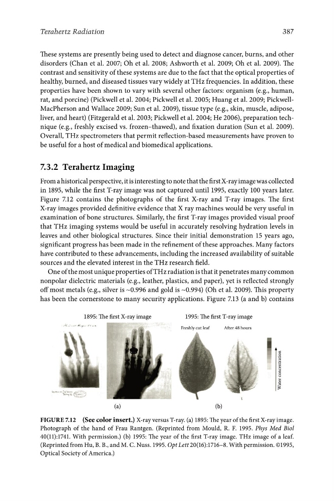
From a historical perspective, it is interesting to note that the first X-ray image was collected
in 1895, while the first T-ray image was not captured until 1995, exactly 100 years later.
Figure 7.12 contains the photographs of the first X-ray and T-ray images. The first
X-ray images provided definitive evidence that X ray machines would be very useful in
examination of bone structures. Similarly, the first T-ray images provided visual proof
that THz imaging systems would be useful in accurately resolving hydration levels in
leaves and other biological structures. Since their initial demonstration 15 years ago,
significant progress has been made in the refinement of these approaches. Many factors
have contributed to these advancements, including the increased availability of suitable
sources and the elevated interest in the THz research field.
One of the most unique properties of THz radiation is that it penetrates many common
nonpolar dielectric materials (e.g., leather, plastics, and paper), yet is reflected strongly
off most metals (e.g., silver is ∼0.996 and gold is ∼0.994) (Oh et al. 2009). This property
has been the cornerstone to many security applications. Figure 7.13 (a and b) contains
Figure 7.12 (See color insert.) X-ray versus T-ray. (a) 1895: The year of the first X-ray image.
Photograph of the hand of Frau Rantgen. (Reprinted from Mould, R. F. 1995. Phys Med Biol
40(11):1741. With permission.) (b) 1995: The year of the first T-ray image. THz image of a leaf.
(Reprinted from Hu, B. B., and M. C. Nuss. 1995. Opt Lett 20(16):1716–8. With permission. ©1995,
Optical Society of America.)
1995: The first T-ray image
Freshly cut leaf
After 48 hours
Water concentration
(a)
(b)
387
Terahertz Radiation
These systems are presently being used to detect and diagnose cancer, burns, and other
disorders (Chan et al. 2007; Oh et al. 2008; Ashworth et al. 2009; Oh et al. 2009). The
contrast and sensitivity of these systems are due to the fact that the optical properties of
healthy, burned, and diseased tissues vary widely at THz frequencies. In addition, these
properties have been shown to vary with several other factors: organism (e.g., human,
rat, and porcine) (Pickwell et al. 2004; Pickwell et al. 2005; Huang et al. 2009; PickwellMacPherson and Wallace 2009; Sun et al. 2009), tissue type (e.g., skin, muscle, adipose,
liver, and heart) (Fitzgerald et al. 2003; Pickwell et al. 2004; He 2006), preparation technique (e.g., freshly excised vs. frozen–thawed), and fixation duration (Sun et al. 2009).
Overall, THz spectrometers that permit reflection-based measurements have proven to
be useful for a host of medical and biomedical applications.
7.3.2 Terahertz Imaging
From a historical perspective, it is interesting to note that the first X-ray image was collected
in 1895, while the first T-ray image was not captured until 1995, exactly 100 years later.
Figure 7.12 contains the photographs of the first X-ray and T-ray images. The first
X-ray images provided definitive evidence that X ray machines would be very useful in
examination of bone structures. Similarly, the first T-ray images provided visual proof
that THz imaging systems would be useful in accurately resolving hydration levels in
leaves and other biological structures. Since their initial demonstration 15 years ago,
significant progress has been made in the refinement of these approaches. Many factors
have contributed to these advancements, including the increased availability of suitable
sources and the elevated interest in the THz research field.
One of the most unique properties of THz radiation is that it penetrates many common
nonpolar dielectric materials (e.g., leather, plastics, and paper), yet is reflected strongly
off most metals (e.g., silver is ∼0.996 and gold is ∼0.994) (Oh et al. 2009). This property
has been the cornerstone to many security applications. Figure 7.13 (a and b) contains
Figure 7.12 (See color insert.) X-ray versus T-ray. (a) 1895: The year of the first X-ray image.
Photograph of the hand of Frau Rantgen. (Reprinted from Mould, R. F. 1995. Phys Med Biol
40(11):1741. With permission.) (b) 1995: The year of the first T-ray image. THz image of a leaf.
(Reprinted from Hu, B. B., and M. C. Nuss. 1995. Opt Lett 20(16):1716–8. With permission. ©1995,
Optical Society of America.)