

(a)
(b)
(c)
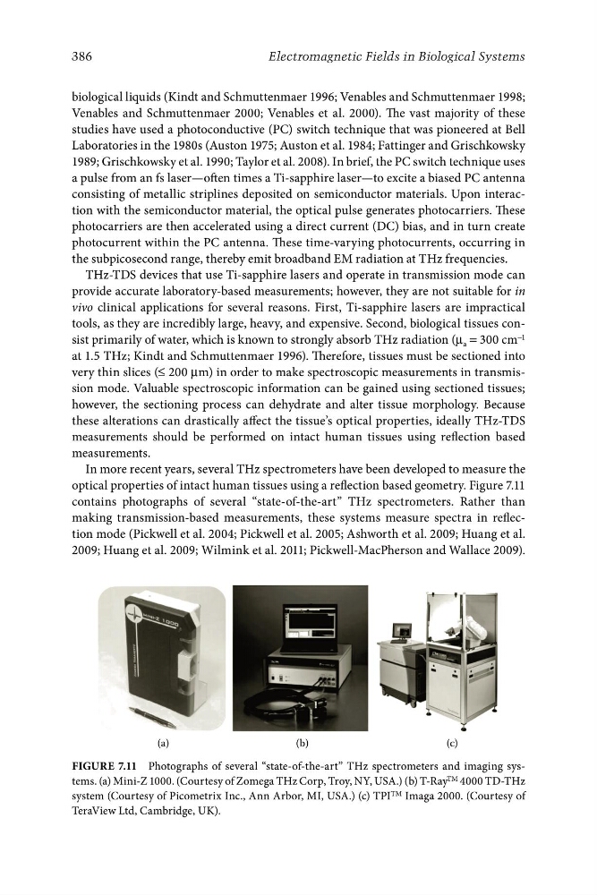
Figure 7.11 Photographs of several “state-of-the-art” THz spectrometers and imaging systems. (a) Mini-Z 1000. (Courtesy of Zomega THz Corp, Troy, NY, USA.) (b) T-Ray TM 4000 TD-THz
system (Courtesy of Picometrix Inc., Ann Arbor, MI, USA.) (c) TPI TM Imaga 2000. (Courtesy of
TeraView Ltd, Cambridge, UK).
386
Electromagnetic Fields in Biological Systems
biological liquids (Kindt and Schmuttenmaer 1996; Venables and Schmuttenmaer 1998;
Venables and Schmuttenmaer 2000; Venables et al. 2000). The vast majority of these
studies have used a photoconductive (PC) switch technique that was pioneered at Bell
Laboratories in the 1980s (Auston 1975; Auston et al. 1984; Fattinger and Grischkowsky
1989; Grischkowsky et al. 1990; Taylor et al. 2008). In brief, the PC switch technique uses
a pulse from an fs laser—often times a Ti-sapphire laser—to excite a biased PC antenna
consisting of metallic striplines deposited on semiconductor materials. Upon interaction with the semiconductor material, the optical pulse generates photocarriers. These
photocarriers are then accelerated using a direct current (DC) bias, and in turn create
photocurrent within the PC antenna. These time-varying photocurrents, occurring in
the subpicosecond range, thereby emit broadband EM radiation at THz frequencies.
THz-TDS devices that use Ti-sapphire lasers and operate in transmission mode can
provide accurate laboratory-based measurements; however, they are not suitable for in
vivo clinical applications for several reasons. First, Ti-sapphire lasers are impractical
tools, as they are incredibly large, heavy, and expensive. Second, biological tissues consist primarily of water, which is known to strongly absorb THz radiation (μ a = 300 cm −1
at 1.5 THz; Kindt and Schmuttenmaer 1996). Therefore, tissues must be sectioned into
very thin slices (≤ 200 μm) in order to make spectroscopic measurements in transmission mode. Valuable spectroscopic information can be gained using sectioned tissues;
however, the sectioning process can dehydrate and alter tissue morphology. Because
these alterations can drastically affect the tissue’s optical properties, ideally THz-TDS
measurements should be performed on intact human tissues using reflection based
measurements.
In more recent years, several THz spectrometers have been developed to measure the
optical properties of intact human tissues using a reflection based geometry. Figure 7.11
contains photographs of several “state-of-the-art” THz spectrometers. Rather than
making transmission-based measurements, these systems measure spectra in reflection mode (Pickwell et al. 2004; Pickwell et al. 2005; Ashworth et al. 2009; Huang et al.
2009; Huang et al. 2009; Wilmink et al. 2011; Pickwell-MacPherson and Wallace 2009).
(b)
(c)
Figure 7.11 Photographs of several “state-of-the-art” THz spectrometers and imaging systems. (a) Mini-Z 1000. (Courtesy of Zomega THz Corp, Troy, NY, USA.) (b) T-Ray TM 4000 TD-THz
system (Courtesy of Picometrix Inc., Ann Arbor, MI, USA.) (c) TPI TM Imaga 2000. (Courtesy of
TeraView Ltd, Cambridge, UK).
386
Electromagnetic Fields in Biological Systems
biological liquids (Kindt and Schmuttenmaer 1996; Venables and Schmuttenmaer 1998;
Venables and Schmuttenmaer 2000; Venables et al. 2000). The vast majority of these
studies have used a photoconductive (PC) switch technique that was pioneered at Bell
Laboratories in the 1980s (Auston 1975; Auston et al. 1984; Fattinger and Grischkowsky
1989; Grischkowsky et al. 1990; Taylor et al. 2008). In brief, the PC switch technique uses
a pulse from an fs laser—often times a Ti-sapphire laser—to excite a biased PC antenna
consisting of metallic striplines deposited on semiconductor materials. Upon interaction with the semiconductor material, the optical pulse generates photocarriers. These
photocarriers are then accelerated using a direct current (DC) bias, and in turn create
photocurrent within the PC antenna. These time-varying photocurrents, occurring in
the subpicosecond range, thereby emit broadband EM radiation at THz frequencies.
THz-TDS devices that use Ti-sapphire lasers and operate in transmission mode can
provide accurate laboratory-based measurements; however, they are not suitable for in
vivo clinical applications for several reasons. First, Ti-sapphire lasers are impractical
tools, as they are incredibly large, heavy, and expensive. Second, biological tissues consist primarily of water, which is known to strongly absorb THz radiation (μ a = 300 cm −1
at 1.5 THz; Kindt and Schmuttenmaer 1996). Therefore, tissues must be sectioned into
very thin slices (≤ 200 μm) in order to make spectroscopic measurements in transmission mode. Valuable spectroscopic information can be gained using sectioned tissues;
however, the sectioning process can dehydrate and alter tissue morphology. Because
these alterations can drastically affect the tissue’s optical properties, ideally THz-TDS
measurements should be performed on intact human tissues using reflection based
measurements.
In more recent years, several THz spectrometers have been developed to measure the
optical properties of intact human tissues using a reflection based geometry. Figure 7.11
contains photographs of several “state-of-the-art” THz spectrometers. Rather than
making transmission-based measurements, these systems measure spectra in reflection mode (Pickwell et al. 2004; Pickwell et al. 2005; Ashworth et al. 2009; Huang et al.
2009; Huang et al. 2009; Wilmink et al. 2011; Pickwell-MacPherson and Wallace 2009).