

(a)
(a)
(c)
(d)
(b)
(b)
1 cm
2 mm
Polypropylene
Gauze
Skin
4 mm
4 mm
Dimensions (mm)
10
20
0.00
7.02
30
(c)
(d)
d1
d2
n2
n1
x
1.0
0.90
0.80
0.70
0.60
0.50
28.12
21.09
14.05
10 20
30 40 50
Dimensions (mm)
(e)
(f )
mV
388
Electromagnetic Fields in Biological Systems
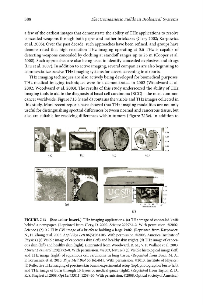
a few of the earliest images that demonstrate the ability of THz applications to resolve
concealed weapons through both paper and leather briefcases (Clery 2002; Karpowicz
et al. 2005). Over the past decade, such approaches have been refined, and groups have
demonstrated that high-resolution THz imaging operating at 0.6 THz is capable of
detecting weapons concealed by clothing at standoff ranges up to 25 m (Cooper et al.
2008). Such approaches are also being used to identify concealed explosives and drugs
(Liu et al. 2007). In addition to active imaging, several companies are also beginning to
commercialize passive THz imaging systems for covert screening in airports.
THz imaging techniques are also actively being developed for biomedical purposes.
THz medical imaging techniques were first demonstrated in 2002 (Woodward et al.
2002; Woodward et al. 2003). The results of this study underscored the ability of THz
imaging tools to aid in the diagnosis of basal cell carcinoma (BCC)—the most common
cancer worldwide. Figure 7.13 (c and d) contains the visible and THz images collected in
this study. More recent reports have showed that THz imaging modalities are not only
useful for distinguishing spectral differences between normal and cancerous tissue, but
also are suitable for resolving differences within tumors (Figure 7.13e). In addition to
Figure 7.13 (See color insert.) THz imaging applications. (a) THz image of concealed knife
behind a newspaper. (Reprinted from Clery, D. 2002. Science 297:761–2. With permission. ©2002,
Science.) (b) 0.2 THz CW image of a briefcase holding a large knife. (Reprinted from Karpowicz,
N., H. Zhong et al. 2005. Appl Phys Lett 86(5):054105. With permission. ©2005, America Institute of
Physics.) (c) Visible image of cancerous skin (left) and healthy skin (right). (d) THz image of cancerous skin (left) and healthy skin (right). (Reprinted from Woodward, R. M., V. P. Wallace et al. 2003.
J Invest Dermatol 120(1):72–8. With permission. ©2003, Nature.) (e) Visible histological image (left)
and THz image (right) of squamous cell carcinoma in lung tissue. (Reprinted from Brun, M. A.,
F. Formanek et al. 2010. Phys Med Biol 55(16):4615. With permission. ©2010, Institute of Physics.)
(f) Reflective THz imaging of porcine skin burns: experimental setup (top), photograph of burn (left),
and THz image of burn through 10 layers of medical gauze (right). (Reprinted from Taylor, Z. D.,
R. S. Singh et al. 2008. Opt Lett 33(11):1258–60. With permission. ©2008, Optical Society of America.)
(a)
(c)
(d)
(b)
(b)
1 cm
2 mm
Polypropylene
Gauze
Skin
4 mm
4 mm
Dimensions (mm)
10
20
0.00
7.02
30
(c)
(d)
d1
d2
n2
n1
x
1.0
0.90
0.80
0.70
0.60
0.50
28.12
21.09
14.05
10 20
30 40 50
Dimensions (mm)
(e)
(f )
mV
388
Electromagnetic Fields in Biological Systems
a few of the earliest images that demonstrate the ability of THz applications to resolve
concealed weapons through both paper and leather briefcases (Clery 2002; Karpowicz
et al. 2005). Over the past decade, such approaches have been refined, and groups have
demonstrated that high-resolution THz imaging operating at 0.6 THz is capable of
detecting weapons concealed by clothing at standoff ranges up to 25 m (Cooper et al.
2008). Such approaches are also being used to identify concealed explosives and drugs
(Liu et al. 2007). In addition to active imaging, several companies are also beginning to
commercialize passive THz imaging systems for covert screening in airports.
THz imaging techniques are also actively being developed for biomedical purposes.
THz medical imaging techniques were first demonstrated in 2002 (Woodward et al.
2002; Woodward et al. 2003). The results of this study underscored the ability of THz
imaging tools to aid in the diagnosis of basal cell carcinoma (BCC)—the most common
cancer worldwide. Figure 7.13 (c and d) contains the visible and THz images collected in
this study. More recent reports have showed that THz imaging modalities are not only
useful for distinguishing spectral differences between normal and cancerous tissue, but
also are suitable for resolving differences within tumors (Figure 7.13e). In addition to
Figure 7.13 (See color insert.) THz imaging applications. (a) THz image of concealed knife
behind a newspaper. (Reprinted from Clery, D. 2002. Science 297:761–2. With permission. ©2002,
Science.) (b) 0.2 THz CW image of a briefcase holding a large knife. (Reprinted from Karpowicz,
N., H. Zhong et al. 2005. Appl Phys Lett 86(5):054105. With permission. ©2005, America Institute of
Physics.) (c) Visible image of cancerous skin (left) and healthy skin (right). (d) THz image of cancerous skin (left) and healthy skin (right). (Reprinted from Woodward, R. M., V. P. Wallace et al. 2003.
J Invest Dermatol 120(1):72–8. With permission. ©2003, Nature.) (e) Visible histological image (left)
and THz image (right) of squamous cell carcinoma in lung tissue. (Reprinted from Brun, M. A.,
F. Formanek et al. 2010. Phys Med Biol 55(16):4615. With permission. ©2010, Institute of Physics.)
(f) Reflective THz imaging of porcine skin burns: experimental setup (top), photograph of burn (left),
and THz image of burn through 10 layers of medical gauze (right). (Reprinted from Taylor, Z. D.,
R. S. Singh et al. 2008. Opt Lett 33(11):1258–60. With permission. ©2008, Optical Society of America.)