

1895: The first X-ray image
1995: The first T-ray image
Freshly cut leaf
After 48 hours
Water concentration
(a)
(b)
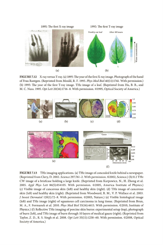
Figure 7.12 X-ray versus T-ray. (a) 1895: The year of the first X-ray image. Photograph of the hand
of Frau Rantgen. (Reprinted from Mould, R. F. 1995. Phys Med Biol 40(11):1741. With permission.)
(b) 1995: The year of the first T-ray image. THz image of a leaf. (Reprinted from Hu, B. B., and
M. C. Nuss. 1995. Opt Lett 20(16):1716–8. With permission. ©1995, Optical Society of America.)
(a)
(a)
(c)
(d)
(b)
(b)
1 cm
2 mm
Polypropylene
Gauze
Skin
4 mm
4 mm
(c)
(d)
Dimensions (mm)
10
20
30
d1
d2
n2
n1
x
1.0
0.90
0.80
0.70
0.60
0.50
28.12
21.09
14.05
7.02
10 20 30 40 50
Dimensions (mm)
(e)
0.00
mV
(f )
Figure 7.13 THz imaging applications. (a) THz image of concealed knife behind a newspaper.
(Reprinted from Clery, D. 2002. Science 297:761–2. With permission. ©2002, Science.) (b) 0.2 THz
CW image of a briefcase holding a large knife. (Reprinted from Karpowicz, N., H. Zhong et al.
2005. Appl Phys Lett 86(5):054105. With permission. ©2005, America Institute of Physics.)
(c) Visible image of cancerous skin (left) and healthy skin (right). (d) THz image of cancerous
skin (left) and healthy skin (right). (Reprinted from Woodward, R. M., V. P. Wallace et al. 2003.
J Invest Dermatol 120(1):72–8. With permission. ©2003, Nature.) (e) Visible histological image
(left) and THz image (right) of squamous cell carcinoma in lung tissue. (Reprinted from Brun,
M. A., F. Formanek et al. 2010. Phys Med Biol 55(16):4615. With permission. ©2010, Institute of
Physics.) (f) Reflective THz imaging of porcine skin burns: experimental setup (top), photograph
of burn (left), and THz image of burn through 10 layers of medical gauze (right). (Reprinted from
Taylor, Z. D., R. S. Singh et al. 2008. Opt Lett 33(11):1258–60. With permission. ©2008, Optical
Society of America.)