

Derivation of cell parameters from measured spectra
Single-shell model
Suspending medium
ε med , σ med
Maxwell–
Wagner
Mixture
model
Single cell
ε cell , σ cell
Cell membrane
ε m , σ m
Cytoplasm
ε cp , σ cp
Cell suspension
ε sus , σ sus
Double-shell model
Nuclear envelope
Nucleoplasm
ε ne , σ ne
ε np , σ np
Spectra simulation with known parameters
103
Pulsed Electric Fields in Biological Cells and Membranes
structural and dynamic parameters of biological tissues (Cole 1975; Cole, Mashimo,
and Winsor 1980; Feldman and Fedotov 1987; Hager 1994). Basically, TDDS is based on
transmission line theory in the time domain that aids in the study of heterogeneities in
coaxial lines according to the change of the shape of a test signal. A rapidly increasing
voltage step V(t) is applied to the line and recorded, along with the reflected voltage R(t)
that is returned from the sample and delayed by the cable propagation time. Any cable
or instrument artifacts are separated from the sample response due to the propagation
delay, thus making them easy to identify and control. The entire frequency spectrum is
captured at once, thus eliminating drift and distortion between frequencies. The complex permittivity is then obtained from these single measurements. An important challenge of this technique though is to correct for electrode polarization effects. Electrode
polarization, which is due to the formation of an electric double layer at the interface
between electrode and conductive biological sample, complicates the determination of
dielectric spectra of cell suspensions and tissues at α- and β-dispersion range. Recent
studies have attempted to address this aspect (Bordi, Cametti, and Gili 2001).
For cell suspensions, the dielectric permittivity ε mix can be expressed by the wellknown Maxwell–Wagner relationship:
ε mix = ε 1 [(2ε 1 + ε c ) − 2p (ε 1 − ε c )]/[(2ε 1 + ε c ) + p (ε 1 − ε c )]
(2.21)
where p is the volume fraction of the suspended cells, ε 1 represents the dielectric permittivity of the liquid phase, and ε c is the cell permittivity. The latter, in turn, can be expressed
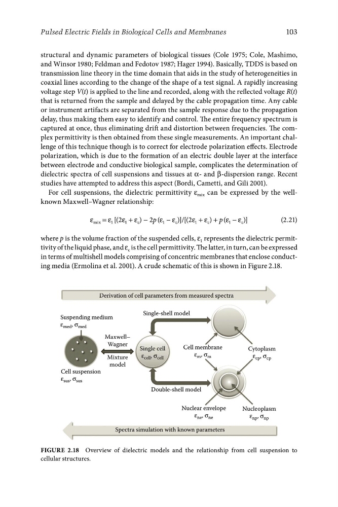
in terms of multishell models comprising of concentric membranes that enclose conducting media (Ermolina et al. 2001). A crude schematic of this is shown in Figure 2.18.
Figure 2.18 Overview of dielectric models and the relationship from cell suspension to
cellular structures.
Single-shell model
Suspending medium
ε med , σ med
Maxwell–
Wagner
Mixture
model
Single cell
ε cell , σ cell
Cell membrane
ε m , σ m
Cytoplasm
ε cp , σ cp
Cell suspension
ε sus , σ sus
Double-shell model
Nuclear envelope
Nucleoplasm
ε ne , σ ne
ε np , σ np
Spectra simulation with known parameters
103
Pulsed Electric Fields in Biological Cells and Membranes
structural and dynamic parameters of biological tissues (Cole 1975; Cole, Mashimo,
and Winsor 1980; Feldman and Fedotov 1987; Hager 1994). Basically, TDDS is based on
transmission line theory in the time domain that aids in the study of heterogeneities in
coaxial lines according to the change of the shape of a test signal. A rapidly increasing
voltage step V(t) is applied to the line and recorded, along with the reflected voltage R(t)
that is returned from the sample and delayed by the cable propagation time. Any cable
or instrument artifacts are separated from the sample response due to the propagation
delay, thus making them easy to identify and control. The entire frequency spectrum is
captured at once, thus eliminating drift and distortion between frequencies. The complex permittivity is then obtained from these single measurements. An important challenge of this technique though is to correct for electrode polarization effects. Electrode
polarization, which is due to the formation of an electric double layer at the interface
between electrode and conductive biological sample, complicates the determination of
dielectric spectra of cell suspensions and tissues at α- and β-dispersion range. Recent
studies have attempted to address this aspect (Bordi, Cametti, and Gili 2001).
For cell suspensions, the dielectric permittivity ε mix can be expressed by the wellknown Maxwell–Wagner relationship:
ε mix = ε 1 [(2ε 1 + ε c ) − 2p (ε 1 − ε c )]/[(2ε 1 + ε c ) + p (ε 1 − ε c )]
(2.21)
where p is the volume fraction of the suspended cells, ε 1 represents the dielectric permittivity of the liquid phase, and ε c is the cell permittivity. The latter, in turn, can be expressed
in terms of multishell models comprising of concentric membranes that enclose conducting media (Ermolina et al. 2001). A crude schematic of this is shown in Figure 2.18.
Figure 2.18 Overview of dielectric models and the relationship from cell suspension to
cellular structures.