

elements lying on their cell surfaces (Raveendran et al. 2018). So far, different fungi
have been deployed for the purpose of synthesizing metal nanoparticles such as
silver, gold, titanium dioxide, and zinc oxide. For example, Krishna et al., in 2017,
exemplified the biogenic synthesis of AgNPs from two white rot fungal strains,
namely, Trametes ljubarskyi and Ganoderma enigmaticum. The results obtained
from their study clearly evidenced that proteins were accountable for the stabilization of AgNPs synthesized by using the cell-free filtrate of Trametes ljubarskyi and
Ganoderma enigmaticum. Correspondingly, Rajput et al. (2016) investigated different fungal strains of Fusarium oxysporum for AgNP synthesis and explored the
effect of isolate selection, temperature, and pH on the morphology of nanoparticles.
In contrast, Molnár et al., in 2018, illustrated the synthesis of AuNPs by employing
29 types of thermophilic fungal strains fermented on different growth media (PDB
and modified Czapek-Dox). The main outcome of this study was that synthesis of
AuNPs can be done via chemical compounds derived from fungi. Yet, one should be
very cautious when the mycelia would be processed further, because the effect of the
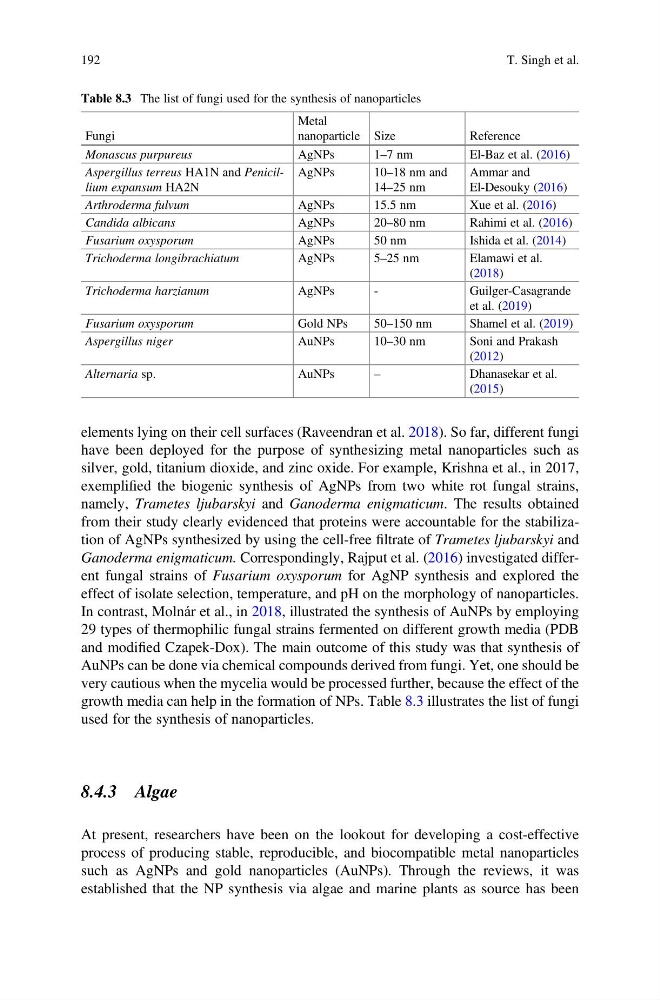
growth media can help in the formation of NPs. Table 8.3 illustrates the list of fungi
used for the synthesis of nanoparticles.
8.4.3 Algae
At present, researchers have been on the lookout for developing a cost-effective
process of producing stable, reproducible, and biocompatible metal nanoparticles
such as AgNPs and gold nanoparticles (AuNPs). Through the reviews, it was
established that the NP synthesis via algae and marine plants as source has been
Table 8.3 The list of fungi used for the synthesis of nanoparticles
Fungi
Metal
nanoparticle Size
Reference
Monascus purpureus
AgNPs
1–7 nm
El-Baz et al. (2016)
Aspergillus terreus HA1N and Penicillium expansum HA2N
AgNPs
10–18 nm and
14–25 nm
Ammar and
El-Desouky (2016)
Arthroderma fulvum
AgNPs
15.5 nm
Xue et al. (2016)
Candida albicans
AgNPs
20–80 nm
Rahimi et al. (2016)
Fusarium oxysporum
AgNPs
50 nm
Ishida et al. (2014)
Trichoderma longibrachiatum
AgNPs
5–25 nm
Elamawi et al.
(2018)
Trichoderma harzianum
AgNPs
-
Guilger-Casagrande
et al. (2019)
Fusarium oxysporum
Gold NPs
50–150 nm
Shamel et al. (2019)
Aspergillus niger
AuNPs
10–30 nm
Soni and Prakash
(2012)
Alternaria sp.
AuNPs
–
Dhanasekar et al.
(2015)
192
T. Singh et al.
have been deployed for the purpose of synthesizing metal nanoparticles such as
silver, gold, titanium dioxide, and zinc oxide. For example, Krishna et al., in 2017,
exemplified the biogenic synthesis of AgNPs from two white rot fungal strains,
namely, Trametes ljubarskyi and Ganoderma enigmaticum. The results obtained
from their study clearly evidenced that proteins were accountable for the stabilization of AgNPs synthesized by using the cell-free filtrate of Trametes ljubarskyi and
Ganoderma enigmaticum. Correspondingly, Rajput et al. (2016) investigated different fungal strains of Fusarium oxysporum for AgNP synthesis and explored the
effect of isolate selection, temperature, and pH on the morphology of nanoparticles.
In contrast, Molnár et al., in 2018, illustrated the synthesis of AuNPs by employing
29 types of thermophilic fungal strains fermented on different growth media (PDB
and modified Czapek-Dox). The main outcome of this study was that synthesis of
AuNPs can be done via chemical compounds derived from fungi. Yet, one should be
very cautious when the mycelia would be processed further, because the effect of the
growth media can help in the formation of NPs. Table 8.3 illustrates the list of fungi
used for the synthesis of nanoparticles.
8.4.3 Algae
At present, researchers have been on the lookout for developing a cost-effective
process of producing stable, reproducible, and biocompatible metal nanoparticles
such as AgNPs and gold nanoparticles (AuNPs). Through the reviews, it was
established that the NP synthesis via algae and marine plants as source has been
Table 8.3 The list of fungi used for the synthesis of nanoparticles
Fungi
Metal
nanoparticle Size
Reference
Monascus purpureus
AgNPs
1–7 nm
El-Baz et al. (2016)
Aspergillus terreus HA1N and Penicillium expansum HA2N
AgNPs
10–18 nm and
14–25 nm
Ammar and
El-Desouky (2016)
Arthroderma fulvum
AgNPs
15.5 nm
Xue et al. (2016)
Candida albicans
AgNPs
20–80 nm
Rahimi et al. (2016)
Fusarium oxysporum
AgNPs
50 nm
Ishida et al. (2014)
Trichoderma longibrachiatum
AgNPs
5–25 nm
Elamawi et al.
(2018)
Trichoderma harzianum
AgNPs
-
Guilger-Casagrande
et al. (2019)
Fusarium oxysporum
Gold NPs
50–150 nm
Shamel et al. (2019)
Aspergillus niger
AuNPs
10–30 nm
Soni and Prakash
(2012)
Alternaria sp.
AuNPs
–
Dhanasekar et al.
(2015)
192
T. Singh et al.