

industries. In 2014, synthesis of silver nanoparticles (AgNPs) via a bacterial strain
Bacillus sp. (CS 11) isolated through heavy metal-contaminated soil was reported.
The isolate CS 11 exhibited the potential to form AgNPs extracellularly within 24 h
at room temperature. In contrast, Shantkriti and Rani in 2014 reported the biosynthesis of copper nanoparticles (CuNPs) by employing supernatant of cell-free system
of non-pathogenic Pseudomonas fluorescens bacteria. The particle size (average)
was established at 49 nm with spherical and hexagonal shapes. Also, Varshney et al.,
in 2010, illustrated an easy, fast, with less costly synthesis of copper nanoparticles
via the non-pathogenic Pseudomonas stutzeri bacteria. The copper nanoparticles
showed great stability. Thus, the metabolites from Pseudomonas stutzeri produced
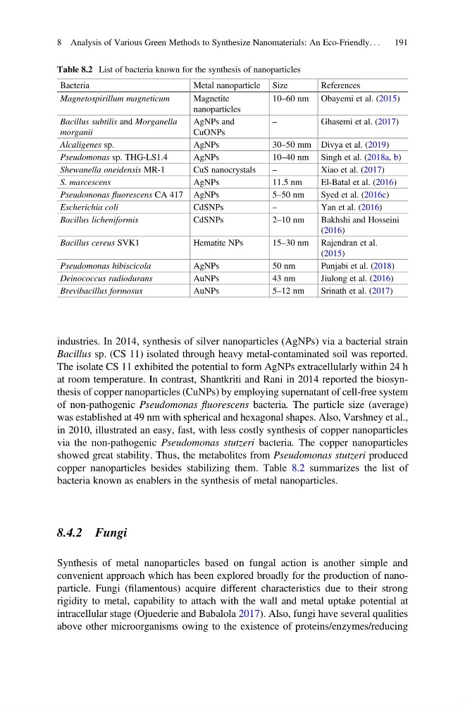
copper nanoparticles besides stabilizing them. Table 8.2 summarizes the list of
bacteria known as enablers in the synthesis of metal nanoparticles.
8.4.2 Fungi
Synthesis of metal nanoparticles based on fungal action is another simple and
convenient approach which has been explored broadly for the production of nanoparticle. Fungi (filamentous) acquire different characteristics due to their strong
rigidity to metal, capability to attach with the wall and metal uptake potential at
intracellular stage (Ojuederie and Babalola 2017). Also, fungi have several qualities
above other microorganisms owing to the existence of proteins/enzymes/reducing
Table 8.2 List of bacteria known for the synthesis of nanoparticles
Bacteria
Metal nanoparticle Size
References
Magnetospirillum magneticum
Magnetite
nanoparticles
10–60 nm Obayemi et al. (2015)
Bacillus subtilis and Morganella
morganii
AgNPs and
CuONPs
–
Ghasemi et al. (2017)
Alcaligenes sp.
AgNPs
30–50 mm Divya et al. (2019)
Pseudomonas sp. THG-LS1.4
AgNPs
10–40 nm Singh et al. (2018a, b)
Shewanella oneidensis MR-1
CuS nanocrystals
–
Xiao et al. (2017)
S. marcescens
AgNPs
11.5 nm
El-Batal et al. (2016)
Pseudomonas fluorescens CA 417 AgNPs
5–50 nm
Syed et al. (2016c)
Escherichia coli
CdSNPs
–
Yan et al. (2016)
Bacillus licheniformis
CdSNPs
2–10 nm
Bakhshi and Hosseini
(2016)
Bacillus cereus SVK1
Hematite NPs
15–30 nm Rajendran et al.
(2015)
Pseudomonas hibiscicola
AgNPs
50 nm
Punjabi et al. (2018)
Deinococcus radiodurans
AuNPs
43 nm
Jiulong et al. (2016)
Brevibacillus formosus
AuNPs
5–12 nm
Srinath et al. (2017)
8 Analysis of Various Green Methods to Synthesize Nanomaterials: An Eco-Friendly. . .
191
Bacillus sp. (CS 11) isolated through heavy metal-contaminated soil was reported.
The isolate CS 11 exhibited the potential to form AgNPs extracellularly within 24 h
at room temperature. In contrast, Shantkriti and Rani in 2014 reported the biosynthesis of copper nanoparticles (CuNPs) by employing supernatant of cell-free system
of non-pathogenic Pseudomonas fluorescens bacteria. The particle size (average)
was established at 49 nm with spherical and hexagonal shapes. Also, Varshney et al.,
in 2010, illustrated an easy, fast, with less costly synthesis of copper nanoparticles
via the non-pathogenic Pseudomonas stutzeri bacteria. The copper nanoparticles
showed great stability. Thus, the metabolites from Pseudomonas stutzeri produced
copper nanoparticles besides stabilizing them. Table 8.2 summarizes the list of
bacteria known as enablers in the synthesis of metal nanoparticles.
8.4.2 Fungi
Synthesis of metal nanoparticles based on fungal action is another simple and
convenient approach which has been explored broadly for the production of nanoparticle. Fungi (filamentous) acquire different characteristics due to their strong
rigidity to metal, capability to attach with the wall and metal uptake potential at
intracellular stage (Ojuederie and Babalola 2017). Also, fungi have several qualities
above other microorganisms owing to the existence of proteins/enzymes/reducing
Table 8.2 List of bacteria known for the synthesis of nanoparticles
Bacteria
Metal nanoparticle Size
References
Magnetospirillum magneticum
Magnetite
nanoparticles
10–60 nm Obayemi et al. (2015)
Bacillus subtilis and Morganella
morganii
AgNPs and
CuONPs
–
Ghasemi et al. (2017)
Alcaligenes sp.
AgNPs
30–50 mm Divya et al. (2019)
Pseudomonas sp. THG-LS1.4
AgNPs
10–40 nm Singh et al. (2018a, b)
Shewanella oneidensis MR-1
CuS nanocrystals
–
Xiao et al. (2017)
S. marcescens
AgNPs
11.5 nm
El-Batal et al. (2016)
Pseudomonas fluorescens CA 417 AgNPs
5–50 nm
Syed et al. (2016c)
Escherichia coli
CdSNPs
–
Yan et al. (2016)
Bacillus licheniformis
CdSNPs
2–10 nm
Bakhshi and Hosseini
(2016)
Bacillus cereus SVK1
Hematite NPs
15–30 nm Rajendran et al.
(2015)
Pseudomonas hibiscicola
AgNPs
50 nm
Punjabi et al. (2018)
Deinococcus radiodurans
AuNPs
43 nm
Jiulong et al. (2016)
Brevibacillus formosus
AuNPs
5–12 nm
Srinath et al. (2017)
8 Analysis of Various Green Methods to Synthesize Nanomaterials: An Eco-Friendly. . .
191