

4.2 Particulate Silica Dispersion as Revealed by 3D-TEM
61
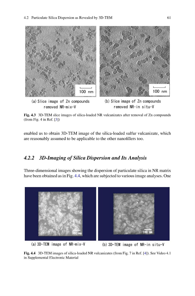
Fig. 4.3 3D-TEM slice images of silica-loaded NR vulcanizates after removal of Zn compounds
(from Fig. 4 in Ref. [3])
enabled us to obtain 3D-TEM image of the silica-loaded sulfur vulcanizate, which
are reasonably assumed to be applicable to the other nanofillers too.
4.2.2 3D-Imaging of Silica Dispersion and Its Analysis
Three-dimensional images showing the dispersion of particulate silica in NR matrix
have been obtained as in Fig. 4.4, which are subjected to various image analyses. One
Fig. 4.4 3D-TEM images of silica-loaded NR vulcanizates (from Fig. 7 in Ref. [4]). See Video 4.1
in Supplemental Electronic Material
61
Fig. 4.3 3D-TEM slice images of silica-loaded NR vulcanizates after removal of Zn compounds
(from Fig. 4 in Ref. [3])
enabled us to obtain 3D-TEM image of the silica-loaded sulfur vulcanizate, which
are reasonably assumed to be applicable to the other nanofillers too.
4.2.2 3D-Imaging of Silica Dispersion and Its Analysis
Three-dimensional images showing the dispersion of particulate silica in NR matrix
have been obtained as in Fig. 4.4, which are subjected to various image analyses. One
Fig. 4.4 3D-TEM images of silica-loaded NR vulcanizates (from Fig. 7 in Ref. [4]). See Video 4.1
in Supplemental Electronic Material