

4.2 Particulate Silica Dispersion as Revealed by 3D-TEM
59
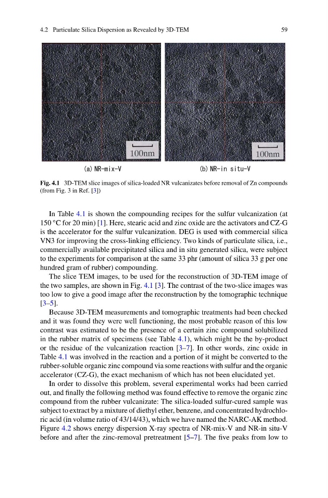
Fig. 4.1 3D-TEM slice images of silica-loaded NR vulcanizates before removal of Zn compounds
(from Fig. 3 in Ref. [3])
In Table 4.1 is shown the compounding recipes for the sulfur vulcanization (at
150 °C for 20 min) [1]. Here, stearic acid and zinc oxide are the activators and CZ-G
is the accelerator for the sulfur vulcanization. DEG is used with commercial silica
VN3 for improving the cross-linking efficiency. Two kinds of particulate silica, i.e.,
commercially available precipitated silica and in situ generated silica, were subject
to the experiments for comparison at the same 33 phr (amount of silica 33 g per one
hundred gram of rubber) compounding.
The slice TEM images, to be used for the reconstruction of 3D-TEM image of
the two samples, are shown in Fig. 4.1 [3]. The contrast of the two-slice images was
too low to give a good image after the reconstruction by the tomographic technique
[3–5].
Because 3D-TEM measurements and tomographic treatments had been checked
and it was found they were well functioning, the most probable reason of this low
contrast was estimated to be the presence of a certain zinc compound solubilized
in the rubber matrix of specimens (see Table 4.1), which might be the by-product
or the residue of the vulcanization reaction [3–7]. In other words, zinc oxide in
Table 4.1 was involved in the reaction and a portion of it might be converted to the
rubber-soluble organic zinc compound via some reactions with sulfur and the organic
accelerator (CZ-G), the exact mechanism of which has not been elucidated yet.
In order to dissolve this problem, several experimental works had been carried
out, and finally the following method was found effective to remove the organic zinc
compound from the rubber vulcanizate: The silica-loaded sulfur-cured sample was
subject to extract by a mixture of diethyl ether, benzene, and concentrated hydrochloric acid (in volume ratio of 43/14/43), which we have named the NARC-AK method.
Figure 4.2 shows energy dispersion X-ray spectra of NR-mix-V and NR-in situ-V
before and after the zinc-removal pretreatment [5–7]. The five peaks from low to
59
Fig. 4.1 3D-TEM slice images of silica-loaded NR vulcanizates before removal of Zn compounds
(from Fig. 3 in Ref. [3])
In Table 4.1 is shown the compounding recipes for the sulfur vulcanization (at
150 °C for 20 min) [1]. Here, stearic acid and zinc oxide are the activators and CZ-G
is the accelerator for the sulfur vulcanization. DEG is used with commercial silica
VN3 for improving the cross-linking efficiency. Two kinds of particulate silica, i.e.,
commercially available precipitated silica and in situ generated silica, were subject
to the experiments for comparison at the same 33 phr (amount of silica 33 g per one
hundred gram of rubber) compounding.
The slice TEM images, to be used for the reconstruction of 3D-TEM image of
the two samples, are shown in Fig. 4.1 [3]. The contrast of the two-slice images was
too low to give a good image after the reconstruction by the tomographic technique
[3–5].
Because 3D-TEM measurements and tomographic treatments had been checked
and it was found they were well functioning, the most probable reason of this low
contrast was estimated to be the presence of a certain zinc compound solubilized
in the rubber matrix of specimens (see Table 4.1), which might be the by-product
or the residue of the vulcanization reaction [3–7]. In other words, zinc oxide in
Table 4.1 was involved in the reaction and a portion of it might be converted to the
rubber-soluble organic zinc compound via some reactions with sulfur and the organic
accelerator (CZ-G), the exact mechanism of which has not been elucidated yet.
In order to dissolve this problem, several experimental works had been carried
out, and finally the following method was found effective to remove the organic zinc
compound from the rubber vulcanizate: The silica-loaded sulfur-cured sample was
subject to extract by a mixture of diethyl ether, benzene, and concentrated hydrochloric acid (in volume ratio of 43/14/43), which we have named the NARC-AK method.
Figure 4.2 shows energy dispersion X-ray spectra of NR-mix-V and NR-in situ-V
before and after the zinc-removal pretreatment [5–7]. The five peaks from low to