

251
9
fraction (V f ), and cracks perpendicular to the beam using conventional radiography. Radiography is not suitable for the evaluation of
the volume fraction (V f ) because it cannot provide accurate data on
the mass or volume of the reinforcement. Also, radiography is not
suitable for detection of interlaminar defects because the dimensions of these defects are very small (in microns) and the reinforcement fibres have high radiation absorption. X-ray microtomography
is another promising NDT technique in which the X-ray beam
focuses on 10-micron spot sizes and can detect interlaminar defects.
9.4.2 Ultrasonic Testing
There are two basic methods for ultrasonic testing: pulse echo and
through-transmission tests. In the pulse-echo transmission method,
the acoustic energy is transmitted through the solid section of the
composite. An ultrasonic probe is placed on one surface or the
outer surface of the composite. A good surface finish of the composite is required so that the probe makes good contact with the
composite. If the acoustic energy encounters any flaw on its travel
path in the form of delamination, voids, or air gaps, the ultrasonic
signal is reflected back.
The principle of the pulse-echo technique is shown in . Fig. 9.16.
In this technique, an ultrasonic wave is transmitted through the
material. A defect in the material reduces the amplitude of the
reflected wave. The depth of the defect is determined by the ratio
D/E p. In the presence of air or a void, the ultrasonic signal will be
reflected back early without travelling through the full thickness of
the composite. That’s how the comparative assessment of the presRadiation source
Specimen
Void
Film
Plan view of the film
Darkerr areas (after processing)
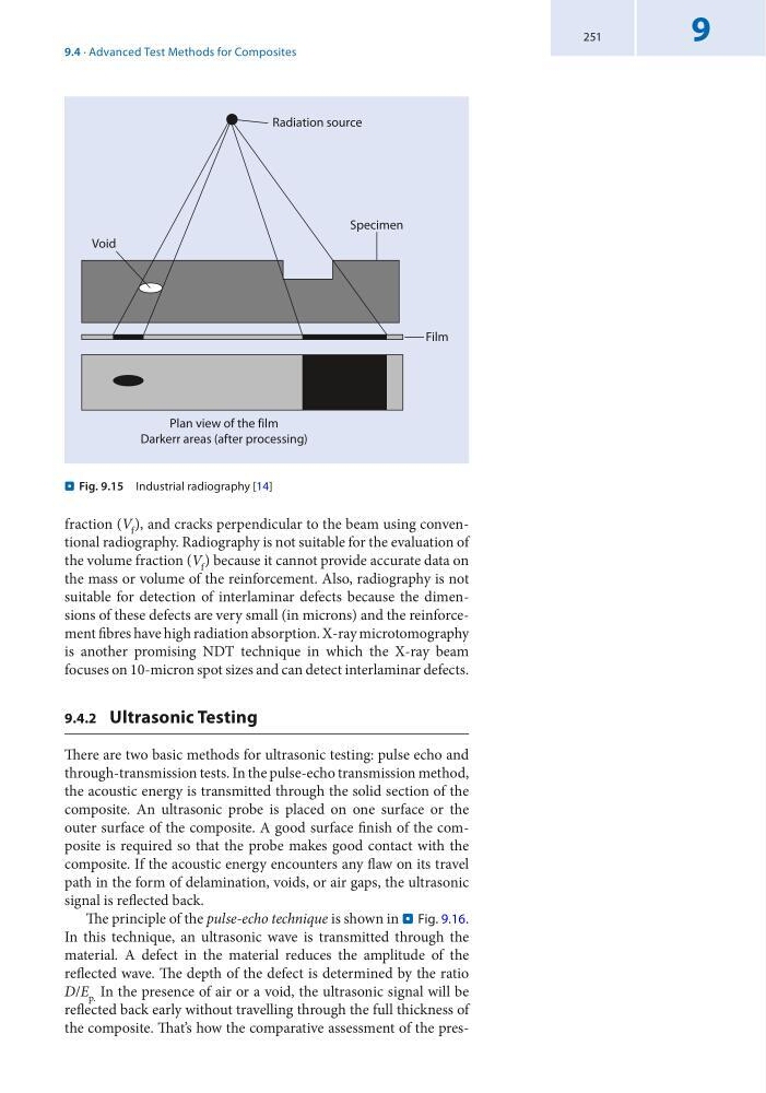
. Fig. 9.15 Industrial radiography [14]
9.4 · Advanced Test Methods for Composites
9
fraction (V f ), and cracks perpendicular to the beam using conventional radiography. Radiography is not suitable for the evaluation of
the volume fraction (V f ) because it cannot provide accurate data on
the mass or volume of the reinforcement. Also, radiography is not
suitable for detection of interlaminar defects because the dimensions of these defects are very small (in microns) and the reinforcement fibres have high radiation absorption. X-ray microtomography
is another promising NDT technique in which the X-ray beam
focuses on 10-micron spot sizes and can detect interlaminar defects.
9.4.2 Ultrasonic Testing
There are two basic methods for ultrasonic testing: pulse echo and
through-transmission tests. In the pulse-echo transmission method,
the acoustic energy is transmitted through the solid section of the
composite. An ultrasonic probe is placed on one surface or the
outer surface of the composite. A good surface finish of the composite is required so that the probe makes good contact with the
composite. If the acoustic energy encounters any flaw on its travel
path in the form of delamination, voids, or air gaps, the ultrasonic
signal is reflected back.
The principle of the pulse-echo technique is shown in . Fig. 9.16.
In this technique, an ultrasonic wave is transmitted through the
material. A defect in the material reduces the amplitude of the
reflected wave. The depth of the defect is determined by the ratio
D/E p. In the presence of air or a void, the ultrasonic signal will be
reflected back early without travelling through the full thickness of
the composite. That’s how the comparative assessment of the presRadiation source
Specimen
Void
Film
Plan view of the film
Darkerr areas (after processing)
. Fig. 9.15 Industrial radiography [14]
9.4 · Advanced Test Methods for Composites