

237
9
3. Transmission Electron Microscopy (TEM)—In TEM, a beam
of electrons is transmitted through an ultra-thin specimen
so that an image is formed from the interaction of the
electrons transmitted through the specimen. The sample
preparation is a specialized activity in TEM. In a sample
preparation, a microtome is used to cut the sample and coat
its surface with carbon. It helps the electron beam to travel
and scan below the surface of the sample without creating a
reflection outside the surface. The depth of penetration is
high in TEM, and it provides information on morphology
even below the surface of the composite. TEM provides finer
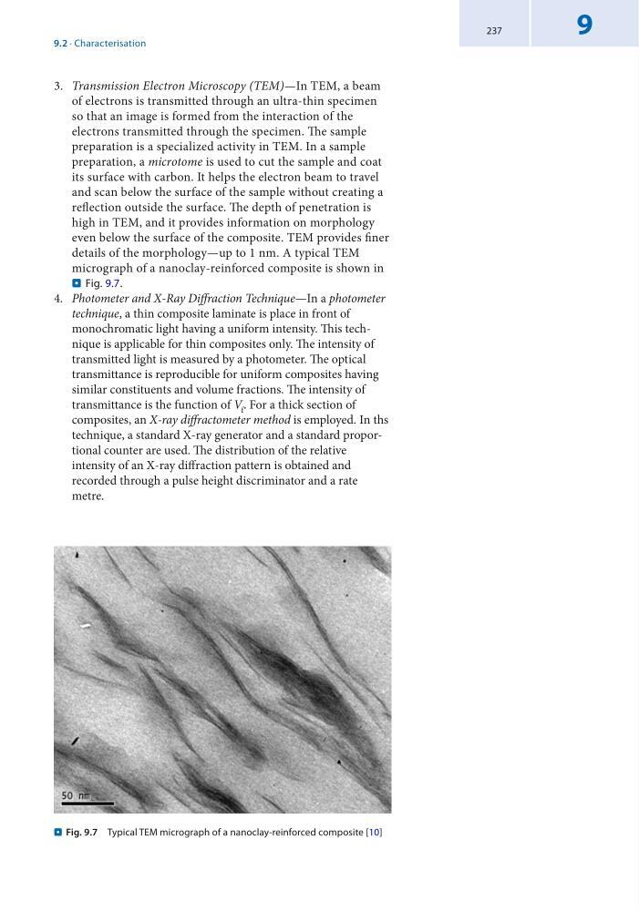
details of the morphology—up to 1 nm. A typical TEM
micrograph of a nanoclay-reinforced composite is shown in
. Fig. 9.7.
4. Photometer and X-Ray Diffraction Technique—In a photometer
technique, a thin composite laminate is place in front of
monochromatic light having a uniform intensity. This technique is applicable for thin composites only. The intensity of
transmitted light is measured by a photometer. The optical
transmittance is reproducible for uniform composites having
similar constituents and volume fractions. The intensity of
transmittance is the function of V f . For a thick section of
composites, an X-ray diffractometer method is employed. In ths
technique, a standard X-ray generator and a standard proportional counter are used. The distribution of the relative
intensity of an X-ray diffraction pattern is obtained and
recorded through a pulse height discriminator and a rate
metre.
. Fig. 9.7 Typical TEM micrograph of a nanoclay-reinforced composite [10]
9.2 · Characterisation
9
3. Transmission Electron Microscopy (TEM)—In TEM, a beam
of electrons is transmitted through an ultra-thin specimen
so that an image is formed from the interaction of the
electrons transmitted through the specimen. The sample
preparation is a specialized activity in TEM. In a sample
preparation, a microtome is used to cut the sample and coat
its surface with carbon. It helps the electron beam to travel
and scan below the surface of the sample without creating a
reflection outside the surface. The depth of penetration is
high in TEM, and it provides information on morphology
even below the surface of the composite. TEM provides finer
details of the morphology—up to 1 nm. A typical TEM
micrograph of a nanoclay-reinforced composite is shown in
. Fig. 9.7.
4. Photometer and X-Ray Diffraction Technique—In a photometer
technique, a thin composite laminate is place in front of
monochromatic light having a uniform intensity. This technique is applicable for thin composites only. The intensity of
transmitted light is measured by a photometer. The optical
transmittance is reproducible for uniform composites having
similar constituents and volume fractions. The intensity of
transmittance is the function of V f . For a thick section of
composites, an X-ray diffractometer method is employed. In ths
technique, a standard X-ray generator and a standard proportional counter are used. The distribution of the relative
intensity of an X-ray diffraction pattern is obtained and
recorded through a pulse height discriminator and a rate
metre.
. Fig. 9.7 Typical TEM micrograph of a nanoclay-reinforced composite [10]
9.2 · Characterisation