

Topics in Current Chemistry (2020) 378:2
1 3
duration (0.5–8 h), leading to different shades of blackness. At the first step of synthesis, titanium sulfate [Ti(SO 4 ) 2 ] and ammonia water were inserted in aqueous
phase inside an ice-water bath in order to control the reaction rate (2 h, under magnetic stirring). After centrifugation and US washing (25 kHz, 100 W, 20 min) by
deionized water, the dispersion was treated with high-power US irradiation (25 kHz,
1500 W/100 mL) using a probe. The synthesis was conducted at 80 °C under different US irradiation durations; 0.5, 1, 2, 4, and 8 h. Afterward, the obtained materials
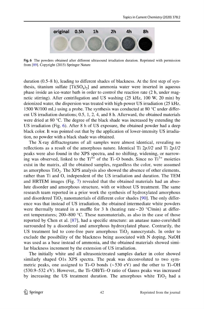
were dried at 80 °C. The degree of the black shade was increased by extending the
US irradiation (Fig. 6). After 8 h of US exposure, the obtained powder had a deep
black color. It was pointed out that by the application of lower-intensity US irradiation, no powder with a black shade was obtained.
The X-ray diffractograms of all samples were almost identical, revealing no
reflections as a result of the amorphous nature. Identical Ti 2p3/2 and Ti 2p1/2
peaks were also found in the XPS spectra, and no shifting, widening, or narrowing was observed, linked to the Ti
4+
of the Ti–O bonds. Since no Ti
3+
moieties
exist in the matrix, all the obtained samples, regardless the color, were assumed
as amorphous TiO 2 . The XPS analysis also showed the absence of other elements,
rather than Ti and O, independent of the US irradiation and duration. The TEM
and HRTEM images (Fig. 7) revealed that the obtained materials had an absolute disorder and amorphous structure, with or without US treatment. The same
research team reported in a prior work the synthesis of hydroxylated amorphous
and disordered TiO 2 nanomaterials of different color shades [90]. The only difference was that instead of US irradiation, the obtained intermediate white powders
were thermally treated in a muffle for 3 h (heating rate ~ 20 °C/min) at different temperatures; 200–800 °C. These nanomaterials, as also in the case of those
reported by Chen et al. [87], had a specific structure: an anatase nano-core/shell
surrounded by a disordered and amorphous hydroxylated phase. Contrarily, the
US treatment led to core-free pure amorphous TiO 2 nanocrystals. In order to
exclude the possibility of the blackness being associated with N doping, NaOH
was used as a base instead of ammonia, and the obtained materials showed similar blackness increment by the extension of US irradiation.
The initially white and all ultrasonictreated samples darker in color showed
similarly shaped O1s XPS spectra. The peak was deconvoluted to two symmetric peaks, one assigned to Ti–O bonds (~ 530 eV) and the other to Ti–OH
(530.9–532 eV). However,, the Ti–OH/Ti–O ratio of Gauss peaks was increased
by increasing the US treatment duration. The amorphous white TiO 2 had a
Fig. 6 The powders obtained after different ultrasound irradiation duration. Reprinted with permission
from [89]. Copyright (2015) Springer Nature
42
Reprinted from the journal
1 3
duration (0.5–8 h), leading to different shades of blackness. At the first step of synthesis, titanium sulfate [Ti(SO 4 ) 2 ] and ammonia water were inserted in aqueous
phase inside an ice-water bath in order to control the reaction rate (2 h, under magnetic stirring). After centrifugation and US washing (25 kHz, 100 W, 20 min) by
deionized water, the dispersion was treated with high-power US irradiation (25 kHz,
1500 W/100 mL) using a probe. The synthesis was conducted at 80 °C under different US irradiation durations; 0.5, 1, 2, 4, and 8 h. Afterward, the obtained materials
were dried at 80 °C. The degree of the black shade was increased by extending the
US irradiation (Fig. 6). After 8 h of US exposure, the obtained powder had a deep
black color. It was pointed out that by the application of lower-intensity US irradiation, no powder with a black shade was obtained.
The X-ray diffractograms of all samples were almost identical, revealing no
reflections as a result of the amorphous nature. Identical Ti 2p3/2 and Ti 2p1/2
peaks were also found in the XPS spectra, and no shifting, widening, or narrowing was observed, linked to the Ti
4+
of the Ti–O bonds. Since no Ti
3+
moieties
exist in the matrix, all the obtained samples, regardless the color, were assumed
as amorphous TiO 2 . The XPS analysis also showed the absence of other elements,
rather than Ti and O, independent of the US irradiation and duration. The TEM
and HRTEM images (Fig. 7) revealed that the obtained materials had an absolute disorder and amorphous structure, with or without US treatment. The same
research team reported in a prior work the synthesis of hydroxylated amorphous
and disordered TiO 2 nanomaterials of different color shades [90]. The only difference was that instead of US irradiation, the obtained intermediate white powders
were thermally treated in a muffle for 3 h (heating rate ~ 20 °C/min) at different temperatures; 200–800 °C. These nanomaterials, as also in the case of those
reported by Chen et al. [87], had a specific structure: an anatase nano-core/shell
surrounded by a disordered and amorphous hydroxylated phase. Contrarily, the
US treatment led to core-free pure amorphous TiO 2 nanocrystals. In order to
exclude the possibility of the blackness being associated with N doping, NaOH
was used as a base instead of ammonia, and the obtained materials showed similar blackness increment by the extension of US irradiation.
The initially white and all ultrasonictreated samples darker in color showed
similarly shaped O1s XPS spectra. The peak was deconvoluted to two symmetric peaks, one assigned to Ti–O bonds (~ 530 eV) and the other to Ti–OH
(530.9–532 eV). However,, the Ti–OH/Ti–O ratio of Gauss peaks was increased
by increasing the US treatment duration. The amorphous white TiO 2 had a
Fig. 6 The powders obtained after different ultrasound irradiation duration. Reprinted with permission
from [89]. Copyright (2015) Springer Nature
42
Reprinted from the journal