

are reports of the damage generated by exposure to NSAIDs on aquatic organisms,
these are not sufficient to determine the impact of these drugs on the aquatic
environment.
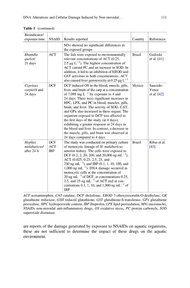
Table 1 (continued)
Bioindicator/
exposure time NSAID Results reported
Country References
MNi showed no significant differences in
the exposed groups
Rhamdia
quelen/
21 days
ACT
The fish were exposed to environmentally
relevant concentrations of ACT (0.25,
2.5 μg L
À1
). The highest concentration of
ACT caused PC and an increase in SOD. In
addition, it led to an inhibition of EROD and
GST activities in both concentrations. ACT
also caused liver genotoxicity at 0.25 μg L
À1
Brazil
Guiloski
et al. [41]
Cyprinus
carpio/4 and
24 days
DCF
DCF induced OS in the blood, muscle, gills,
liver, and brain of the carp at a concentration
of 7.098 mg L
À1 by exposure to 4 and
24 days. There were significant increases in
HPC, LPX, and PC in blood, muscles, gills,
brain, and liver. The activity of SOD, CAT,
and GPx also increased in these organs. The
organism exposed to DCF was affected in
the first days of the study (at 4 days),
exhibiting a greater response at 24 days in
the blood and liver. In contrast, a decrease in
the muscle, gills, and brain was observed at
24 days compared to 4 days
Mexico
SaucedoVence
et al. [42]
Hoplias
malabaricus/
after 24 h
DCF
ACT
IBP
The study was conducted on primary culture
of monocytic lineage of H. malabaricus
anterior kidney. The cells were exposed to
DCF (0.2, 2, 20, 200, and 20,000 ng mL
À1
),
ACT (0.025, 0.25, 2.5, 25, and
250 ng mL
À1
), and IBP (0.1, 1, 10, 100, and
1,000 ng mL
À1
). DNA damage occurred in
monocytic cells at the concentration of
20 ng mL
À1 of DCF, at concentrations 0.25,
2.5, and 25 ng mL
À1 of ACT and at concentrations 0.1, 1, 10, and 1,000 ng mL
À1 of
IBP
Brazil
Ribas et al.
[43]
ACT acetaminophen, CAT catalase, DCF diclofenac, EROD 7-ethoxyresorufin-O-deethylase, GR
glutathione reductase, GSH reduced glutathione, GST glutathione-S-transferase, GPx glutathione
peroxidase, HPC hydroperoxide content, IBP ibuprofen, LPX lipid peroxidation, MNi micronuclei,
NSAIDs non-steroidal anti-inflammatory drugs, OS oxidative stress, PC protein carbonyls, SOD
superoxide dismutase
DNA Alterations and Cellular Damage Induced by Non-steroidal. . .
111
these are not sufficient to determine the impact of these drugs on the aquatic
environment.
Table 1 (continued)
Bioindicator/
exposure time NSAID Results reported
Country References
MNi showed no significant differences in
the exposed groups
Rhamdia
quelen/
21 days
ACT
The fish were exposed to environmentally
relevant concentrations of ACT (0.25,
2.5 μg L
À1
). The highest concentration of
ACT caused PC and an increase in SOD. In
addition, it led to an inhibition of EROD and
GST activities in both concentrations. ACT
also caused liver genotoxicity at 0.25 μg L
À1
Brazil
Guiloski
et al. [41]
Cyprinus
carpio/4 and
24 days
DCF
DCF induced OS in the blood, muscle, gills,
liver, and brain of the carp at a concentration
of 7.098 mg L
À1 by exposure to 4 and
24 days. There were significant increases in
HPC, LPX, and PC in blood, muscles, gills,
brain, and liver. The activity of SOD, CAT,
and GPx also increased in these organs. The
organism exposed to DCF was affected in
the first days of the study (at 4 days),
exhibiting a greater response at 24 days in
the blood and liver. In contrast, a decrease in
the muscle, gills, and brain was observed at
24 days compared to 4 days
Mexico
SaucedoVence
et al. [42]
Hoplias
malabaricus/
after 24 h
DCF
ACT
IBP
The study was conducted on primary culture
of monocytic lineage of H. malabaricus
anterior kidney. The cells were exposed to
DCF (0.2, 2, 20, 200, and 20,000 ng mL
À1
),
ACT (0.025, 0.25, 2.5, 25, and
250 ng mL
À1
), and IBP (0.1, 1, 10, 100, and
1,000 ng mL
À1
). DNA damage occurred in
monocytic cells at the concentration of
20 ng mL
À1 of DCF, at concentrations 0.25,
2.5, and 25 ng mL
À1 of ACT and at concentrations 0.1, 1, 10, and 1,000 ng mL
À1 of
IBP
Brazil
Ribas et al.
[43]
ACT acetaminophen, CAT catalase, DCF diclofenac, EROD 7-ethoxyresorufin-O-deethylase, GR
glutathione reductase, GSH reduced glutathione, GST glutathione-S-transferase, GPx glutathione
peroxidase, HPC hydroperoxide content, IBP ibuprofen, LPX lipid peroxidation, MNi micronuclei,
NSAIDs non-steroidal anti-inflammatory drugs, OS oxidative stress, PC protein carbonyls, SOD
superoxide dismutase
DNA Alterations and Cellular Damage Induced by Non-steroidal. . .
111