

16.3 Results and Discussions
X-ray computed microtomography presents itself as an accurate test for the characterization of the cellular structure of metal
foams. The images that make up Fig. 16.5 were obtained by longitudinal frames, that is, top views of longitudinal sections of
the analyzed sample, with the image (a) representing the solid lower disk of the aluminum alloy, and the (b), (c) and
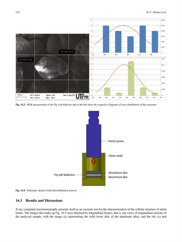
Fig. 16.3 SEM measurement of the Fly Ash balloons and in the left show the respective diagrams of sizes distribution of this measures
Fig. 16.4 Schematic sketch of the thixoinfiltration process
110
R. C. Moraes et al.
X-ray computed microtomography presents itself as an accurate test for the characterization of the cellular structure of metal
foams. The images that make up Fig. 16.5 were obtained by longitudinal frames, that is, top views of longitudinal sections of
the analyzed sample, with the image (a) representing the solid lower disk of the aluminum alloy, and the (b), (c) and
Fig. 16.3 SEM measurement of the Fly Ash balloons and in the left show the respective diagrams of sizes distribution of this measures
Fig. 16.4 Schematic sketch of the thixoinfiltration process
110
R. C. Moraes et al.