

464
8 Morphogenesis
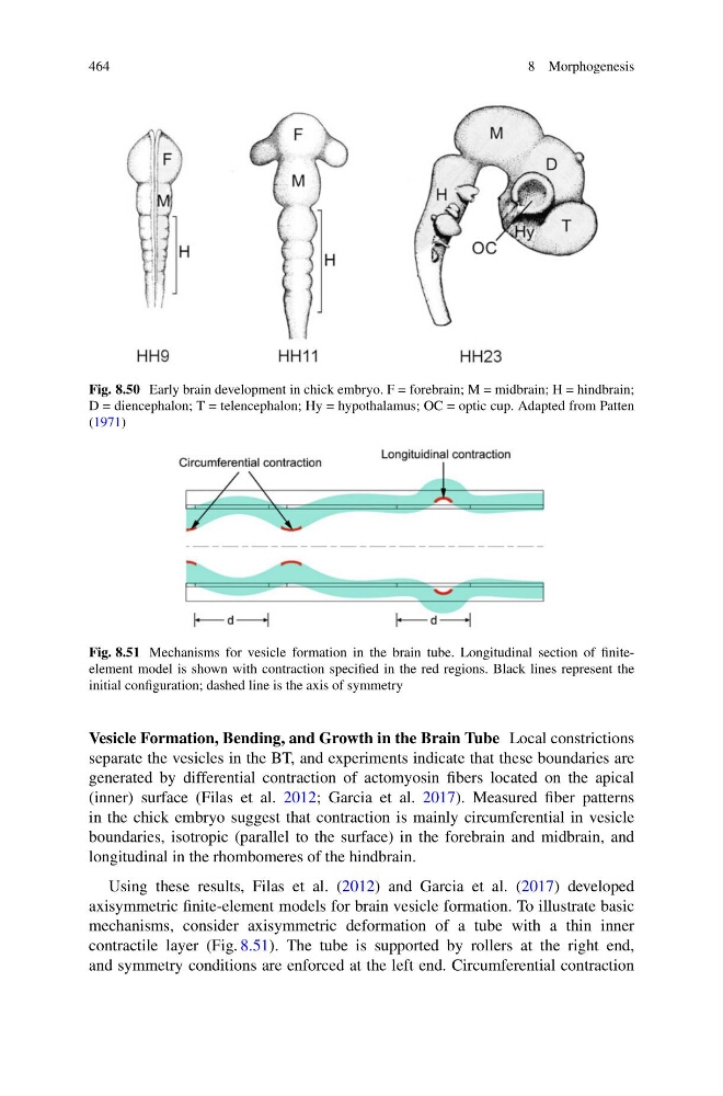
Fig. 8.50 Early brain development in chick embryo. F = forebrain; M = midbrain; H = hindbrain;
D = diencephalon; T = telencephalon; Hy = hypothalamus; OC = optic cup. Adapted from Patten
(1971)
Fig. 8.51 Mechanisms for vesicle formation in the brain tube. Longitudinal section of finiteelement model is shown with contraction specified in the red regions. Black lines represent the
initial configuration; dashed line is the axis of symmetry
Vesicle Formation, Bending, and Growth in the Brain Tube Local constrictions
separate the vesicles in the BT, and experiments indicate that these boundaries are
generated by differential contraction of actomyosin fibers located on the apical
(inner) surface (Filas et al. 2012; Garcia et al. 2017). Measured fiber patterns
in the chick embryo suggest that contraction is mainly circumferential in vesicle
boundaries, isotropic (parallel to the surface) in the forebrain and midbrain, and
longitudinal in the rhombomeres of the hindbrain.
Using these results, Filas et al. (2012) and Garcia et al. (2017) developed
axisymmetric finite-element models for brain vesicle formation. To illustrate basic
mechanisms, consider axisymmetric deformation of a tube with a thin inner
contractile layer (Fig. 8.51). The tube is supported by rollers at the right end,
and symmetry conditions are enforced at the left end. Circumferential contraction
8 Morphogenesis
Fig. 8.50 Early brain development in chick embryo. F = forebrain; M = midbrain; H = hindbrain;
D = diencephalon; T = telencephalon; Hy = hypothalamus; OC = optic cup. Adapted from Patten
(1971)
Fig. 8.51 Mechanisms for vesicle formation in the brain tube. Longitudinal section of finiteelement model is shown with contraction specified in the red regions. Black lines represent the
initial configuration; dashed line is the axis of symmetry
Vesicle Formation, Bending, and Growth in the Brain Tube Local constrictions
separate the vesicles in the BT, and experiments indicate that these boundaries are
generated by differential contraction of actomyosin fibers located on the apical
(inner) surface (Filas et al. 2012; Garcia et al. 2017). Measured fiber patterns
in the chick embryo suggest that contraction is mainly circumferential in vesicle
boundaries, isotropic (parallel to the surface) in the forebrain and midbrain, and
longitudinal in the rhombomeres of the hindbrain.
Using these results, Filas et al. (2012) and Garcia et al. (2017) developed
axisymmetric finite-element models for brain vesicle formation. To illustrate basic
mechanisms, consider axisymmetric deformation of a tube with a thin inner
contractile layer (Fig. 8.51). The tube is supported by rollers at the right end,
and symmetry conditions are enforced at the left end. Circumferential contraction