

8.6 Neural Development
463
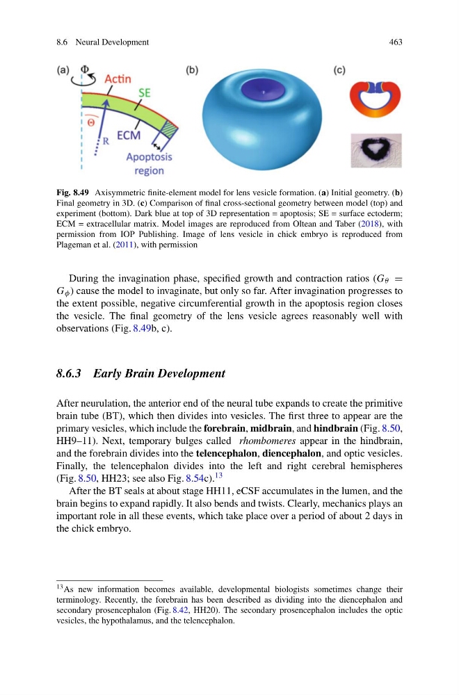
Fig. 8.49 Axisymmetric finite-element model for lens vesicle formation. (a) Initial geometry. (b)
Final geometry in 3D. (c) Comparison of final cross-sectional geometry between model (top) and
experiment (bottom). Dark blue at top of 3D representation = apoptosis; SE = surface ectoderm;
ECM = extracellular matrix. Model images are reproduced from Oltean and Taber (2018), with
permission from IOP Publishing. Image of lens vesicle in chick embryo is reproduced from
Plageman et al. (2011), with permission
During the invagination phase, specified growth and contraction ratios (G θ =
G φ ) cause the model to invaginate, but only so far. After invagination progresses to
the extent possible, negative circumferential growth in the apoptosis region closes
the vesicle. The final geometry of the lens vesicle agrees reasonably well with
observations (Fig. 8.49b, c).
8.6.3 Early Brain Development
After neurulation, the anterior end of the neural tube expands to create the primitive
brain tube (BT), which then divides into vesicles. The first three to appear are the
primary vesicles, which include the forebrain, midbrain, and hindbrain (Fig. 8.50,
HH9–11). Next, temporary bulges called rhombomeres appear in the hindbrain,
and the forebrain divides into the telencephalon, diencephalon, and optic vesicles.
Finally, the telencephalon divides into the left and right cerebral hemispheres
(Fig. 8.50, HH23; see also Fig. 8.54c). 13
After the BT seals at about stage HH11, eCSF accumulates in the lumen, and the
brain begins to expand rapidly. It also bends and twists. Clearly, mechanics plays an
important role in all these events, which take place over a period of about 2 days in
the chick embryo.
13 As new information becomes available, developmental biologists sometimes change their
terminology. Recently, the forebrain has been described as dividing into the diencephalon and
secondary prosencephalon (Fig. 8.42, HH20). The secondary prosencephalon includes the optic
vesicles, the hypothalamus, and the telencephalon.
463
Fig. 8.49 Axisymmetric finite-element model for lens vesicle formation. (a) Initial geometry. (b)
Final geometry in 3D. (c) Comparison of final cross-sectional geometry between model (top) and
experiment (bottom). Dark blue at top of 3D representation = apoptosis; SE = surface ectoderm;
ECM = extracellular matrix. Model images are reproduced from Oltean and Taber (2018), with
permission from IOP Publishing. Image of lens vesicle in chick embryo is reproduced from
Plageman et al. (2011), with permission
During the invagination phase, specified growth and contraction ratios (G θ =
G φ ) cause the model to invaginate, but only so far. After invagination progresses to
the extent possible, negative circumferential growth in the apoptosis region closes
the vesicle. The final geometry of the lens vesicle agrees reasonably well with
observations (Fig. 8.49b, c).
8.6.3 Early Brain Development
After neurulation, the anterior end of the neural tube expands to create the primitive
brain tube (BT), which then divides into vesicles. The first three to appear are the
primary vesicles, which include the forebrain, midbrain, and hindbrain (Fig. 8.50,
HH9–11). Next, temporary bulges called rhombomeres appear in the hindbrain,
and the forebrain divides into the telencephalon, diencephalon, and optic vesicles.
Finally, the telencephalon divides into the left and right cerebral hemispheres
(Fig. 8.50, HH23; see also Fig. 8.54c). 13
After the BT seals at about stage HH11, eCSF accumulates in the lumen, and the
brain begins to expand rapidly. It also bends and twists. Clearly, mechanics plays an
important role in all these events, which take place over a period of about 2 days in
the chick embryo.
13 As new information becomes available, developmental biologists sometimes change their
terminology. Recently, the forebrain has been described as dividing into the diencephalon and
secondary prosencephalon (Fig. 8.42, HH20). The secondary prosencephalon includes the optic
vesicles, the hypothalamus, and the telencephalon.