

448
8 Morphogenesis
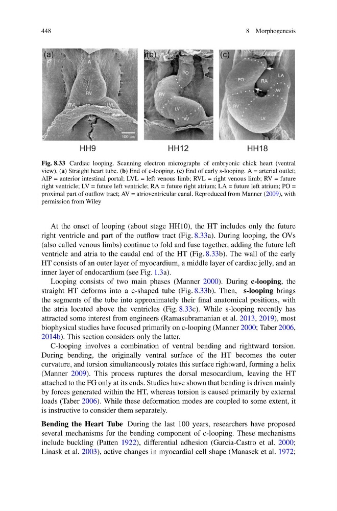
Fig. 8.33 Cardiac looping. Scanning electron micrographs of embryonic chick heart (ventral
view). (a) Straight heart tube. (b) End of c-looping. (c) End of early s-looping. A = arterial outlet;
AIP = anterior intestinal portal; LVL = left venous limb; RVL = right venous limb; RV = future
right ventricle; LV = future left ventricle; RA = future right atrium; LA = future left atrium; PO =
proximal part of outflow tract; AV = atrioventricular canal. Reproduced from Manner (2009), with
permission from Wiley
At the onset of looping (about stage HH10), the HT includes only the future
right ventricle and part of the outflow tract (Fig. 8.33a). During looping, the OVs
(also called venous limbs) continue to fold and fuse together, adding the future left
ventricle and atria to the caudal end of the HT (Fig. 8.33b). The wall of the early
HT consists of an outer layer of myocardium, a middle layer of cardiac jelly, and an
inner layer of endocardium (see Fig. 1.3a).
Looping consists of two main phases (Manner 2000). During c-looping, the
straight HT deforms into a c-shaped tube (Fig. 8.33b). Then, s-looping brings
the segments of the tube into approximately their final anatomical positions, with
the atria located above the ventricles (Fig. 8.33c). While s-looping recently has
attracted some interest from engineers (Ramasubramanian et al. 2013, 2019), most
biophysical studies have focused primarily on c-looping (Manner 2000; Taber 2006,
2014b). This section considers only the latter.
C-looping involves a combination of ventral bending and rightward torsion.
During bending, the originally ventral surface of the HT becomes the outer
curvature, and torsion simultaneously rotates this surface rightward, forming a helix
(Manner 2009). This process ruptures the dorsal mesocardium, leaving the HT
attached to the FG only at its ends. Studies have shown that bending is driven mainly
by forces generated within the HT, whereas torsion is caused primarily by external
loads (Taber 2006). While these deformation modes are coupled to some extent, it
is instructive to consider them separately.
Bending the Heart Tube During the last 100 years, researchers have proposed
several mechanisms for the bending component of c-looping. These mechanisms
include buckling (Patten 1922), differential adhesion (Garcia-Castro et al. 2000;
Linask et al. 2003), active changes in myocardial cell shape (Manasek et al. 1972;
8 Morphogenesis
Fig. 8.33 Cardiac looping. Scanning electron micrographs of embryonic chick heart (ventral
view). (a) Straight heart tube. (b) End of c-looping. (c) End of early s-looping. A = arterial outlet;
AIP = anterior intestinal portal; LVL = left venous limb; RVL = right venous limb; RV = future
right ventricle; LV = future left ventricle; RA = future right atrium; LA = future left atrium; PO =
proximal part of outflow tract; AV = atrioventricular canal. Reproduced from Manner (2009), with
permission from Wiley
At the onset of looping (about stage HH10), the HT includes only the future
right ventricle and part of the outflow tract (Fig. 8.33a). During looping, the OVs
(also called venous limbs) continue to fold and fuse together, adding the future left
ventricle and atria to the caudal end of the HT (Fig. 8.33b). The wall of the early
HT consists of an outer layer of myocardium, a middle layer of cardiac jelly, and an
inner layer of endocardium (see Fig. 1.3a).
Looping consists of two main phases (Manner 2000). During c-looping, the
straight HT deforms into a c-shaped tube (Fig. 8.33b). Then, s-looping brings
the segments of the tube into approximately their final anatomical positions, with
the atria located above the ventricles (Fig. 8.33c). While s-looping recently has
attracted some interest from engineers (Ramasubramanian et al. 2013, 2019), most
biophysical studies have focused primarily on c-looping (Manner 2000; Taber 2006,
2014b). This section considers only the latter.
C-looping involves a combination of ventral bending and rightward torsion.
During bending, the originally ventral surface of the HT becomes the outer
curvature, and torsion simultaneously rotates this surface rightward, forming a helix
(Manner 2009). This process ruptures the dorsal mesocardium, leaving the HT
attached to the FG only at its ends. Studies have shown that bending is driven mainly
by forces generated within the HT, whereas torsion is caused primarily by external
loads (Taber 2006). While these deformation modes are coupled to some extent, it
is instructive to consider them separately.
Bending the Heart Tube During the last 100 years, researchers have proposed
several mechanisms for the bending component of c-looping. These mechanisms
include buckling (Patten 1922), differential adhesion (Garcia-Castro et al. 2000;
Linask et al. 2003), active changes in myocardial cell shape (Manasek et al. 1972;