

8.5 Early Heart Development
447
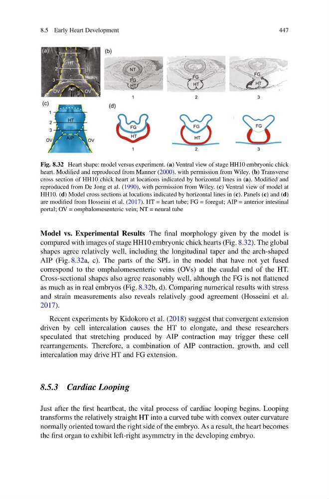
Fig. 8.32 Heart shape: model versus experiment. (a) Ventral view of stage HH10 embryonic chick
heart. Modified and reproduced from Manner (2000), with permission from Wiley. (b) Transverse
cross section of HH10 chick heart at locations indicated by horizontal lines in (a). Modified and
reproduced from De Jong et al. (1990), with permission from Wiley. (c) Ventral view of model at
HH10. (d) Model cross sections at locations indicated by horizontal lines in (c). Panels (c) and (d)
are modified from Hosseini et al. (2017). HT = heart tube; FG = foregut; AIP = anterior intestinal
portal; OV = omphalomesenteric vein; NT = neural tube
Model vs. Experimental Results The final morphology given by the model is
compared with images of stage HH10 embryonic chick hearts (Fig. 8.32). The global
shapes agree relatively well, including the longitudinal taper and the arch-shaped
AIP (Fig. 8.32a, c). The parts of the SPL in the model that have not yet fused
correspond to the omphalomesenteric veins (OVs) at the caudal end of the HT.
Cross-sectional shapes also agree reasonably well, although the FG is not flattened
as much as in real embryos (Fig. 8.32b, d). Comparing numerical results with stress
and strain measurements also reveals relatively good agreement (Hosseini et al.
2017).
Recent experiments by Kidokoro et al. (2018) suggest that convergent extension
driven by cell intercalation causes the HT to elongate, and these researchers
speculated that stretching produced by AIP contraction may trigger these cell
rearrangements. Therefore, a combination of AIP contraction, growth, and cell
intercalation may drive HT and FG extension.
8.5.3 Cardiac Looping
Just after the first heartbeat, the vital process of cardiac looping begins. Looping
transforms the relatively straight HT into a curved tube with convex outer curvature
normally oriented toward the right side of the embryo. As a result, the heart becomes
the first organ to exhibit left-right asymmetry in the developing embryo.
447
Fig. 8.32 Heart shape: model versus experiment. (a) Ventral view of stage HH10 embryonic chick
heart. Modified and reproduced from Manner (2000), with permission from Wiley. (b) Transverse
cross section of HH10 chick heart at locations indicated by horizontal lines in (a). Modified and
reproduced from De Jong et al. (1990), with permission from Wiley. (c) Ventral view of model at
HH10. (d) Model cross sections at locations indicated by horizontal lines in (c). Panels (c) and (d)
are modified from Hosseini et al. (2017). HT = heart tube; FG = foregut; AIP = anterior intestinal
portal; OV = omphalomesenteric vein; NT = neural tube
Model vs. Experimental Results The final morphology given by the model is
compared with images of stage HH10 embryonic chick hearts (Fig. 8.32). The global
shapes agree relatively well, including the longitudinal taper and the arch-shaped
AIP (Fig. 8.32a, c). The parts of the SPL in the model that have not yet fused
correspond to the omphalomesenteric veins (OVs) at the caudal end of the HT.
Cross-sectional shapes also agree reasonably well, although the FG is not flattened
as much as in real embryos (Fig. 8.32b, d). Comparing numerical results with stress
and strain measurements also reveals relatively good agreement (Hosseini et al.
2017).
Recent experiments by Kidokoro et al. (2018) suggest that convergent extension
driven by cell intercalation causes the HT to elongate, and these researchers
speculated that stretching produced by AIP contraction may trigger these cell
rearrangements. Therefore, a combination of AIP contraction, growth, and cell
intercalation may drive HT and FG extension.
8.5.3 Cardiac Looping
Just after the first heartbeat, the vital process of cardiac looping begins. Looping
transforms the relatively straight HT into a curved tube with convex outer curvature
normally oriented toward the right side of the embryo. As a result, the heart becomes
the first organ to exhibit left-right asymmetry in the developing embryo.