

444
8 Morphogenesis
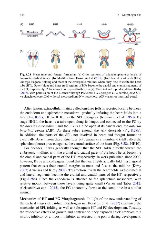
Fig. 8.28 Heart tube and foregut formation. (a) Cross sections of splanchnopleure at levels of
horizontal dashed lines in (b). Modified from Hosseini et al. (2017). (b) Bilateral heart fields (HFs)
undergo diagonal folding and meet at the embryonic midline, where they fuse to create the heart
tube (HT). Outer (blue) and inner (red) regions of HFs become the caudal and cranial segments of
the HT, respectively. Colors do not correspond to those in (a). Modified and reproduced from Kirby
(2007), with permission of the Licensor through PLSclear. FG = foregut; CJ = cardiac jelly; SPL
= splanchnopleure; DM = dorsal mesocardium; N = notochord; AIP = anterior intestinal portal
After fusion, extracellular matrix called cardiac jelly is secreted locally between
the endoderm and splanchnic mesoderm, gradually inflating the heart fields into a
tube (Fig. 8.28a, HH8–HH10), as the SPL elongates (Romanoff et al. 1960). By
stage HH10, the heart is a tube open along its length and connected to the FG by
the dorsal mesocardium; and the FG is a tube open at its caudal end, the anterior
intestinal portal (AIP). As these tubes extend, the AIP descends (Fig. 8.28b).
In addition, the parts of the SPL not involved in heart and foregut formation
eventually detach from these structures but remain as a membrane (still called the
splanchnopleure) pressed against the ventral surface of the heart (Fig. 8.28a, HH10).
For decades, it was generally thought that the SPL folds directly toward the
embryonic midline, with the cranial and caudal parts of the heart fields becoming
the cranial and caudal parts of the HT, respectively. In work published since 2000,
however, Kirby and colleagues found that the heart fields actually fold in a diagonal
pattern that causes their cranial margins to meet and fuse at the midline (Kirby
2007; Abu-Issa and Kirby 2008). This motion inverts the heart fields, as their medial
and lateral segments become the cranial and caudal parts of the HT, respectively
(Fig. 8.28b). Since the endoderm is attached to the splanchnic mesoderm, with
relative motion between these layers being quite small (Varner and Taber 2012;
Aleksandrova et al. 2015), the FG apparently forms at the same time in a similar
manner.
Mechanics of HT and FG Morphogenesis In light of the new understanding of
the earliest stages of cardiac morphogenesis, Hosseini et al. (2017) examined the
mechanics of SPL folding, as well as subsequent HT and FG development. To study
the respective effects of growth and contraction, they exposed chick embryos to a
mitotic inhibitor or a myosin inhibitor at selected time points during development.
8 Morphogenesis
Fig. 8.28 Heart tube and foregut formation. (a) Cross sections of splanchnopleure at levels of
horizontal dashed lines in (b). Modified from Hosseini et al. (2017). (b) Bilateral heart fields (HFs)
undergo diagonal folding and meet at the embryonic midline, where they fuse to create the heart
tube (HT). Outer (blue) and inner (red) regions of HFs become the caudal and cranial segments of
the HT, respectively. Colors do not correspond to those in (a). Modified and reproduced from Kirby
(2007), with permission of the Licensor through PLSclear. FG = foregut; CJ = cardiac jelly; SPL
= splanchnopleure; DM = dorsal mesocardium; N = notochord; AIP = anterior intestinal portal
After fusion, extracellular matrix called cardiac jelly is secreted locally between
the endoderm and splanchnic mesoderm, gradually inflating the heart fields into a
tube (Fig. 8.28a, HH8–HH10), as the SPL elongates (Romanoff et al. 1960). By
stage HH10, the heart is a tube open along its length and connected to the FG by
the dorsal mesocardium; and the FG is a tube open at its caudal end, the anterior
intestinal portal (AIP). As these tubes extend, the AIP descends (Fig. 8.28b).
In addition, the parts of the SPL not involved in heart and foregut formation
eventually detach from these structures but remain as a membrane (still called the
splanchnopleure) pressed against the ventral surface of the heart (Fig. 8.28a, HH10).
For decades, it was generally thought that the SPL folds directly toward the
embryonic midline, with the cranial and caudal parts of the heart fields becoming
the cranial and caudal parts of the HT, respectively. In work published since 2000,
however, Kirby and colleagues found that the heart fields actually fold in a diagonal
pattern that causes their cranial margins to meet and fuse at the midline (Kirby
2007; Abu-Issa and Kirby 2008). This motion inverts the heart fields, as their medial
and lateral segments become the cranial and caudal parts of the HT, respectively
(Fig. 8.28b). Since the endoderm is attached to the splanchnic mesoderm, with
relative motion between these layers being quite small (Varner and Taber 2012;
Aleksandrova et al. 2015), the FG apparently forms at the same time in a similar
manner.
Mechanics of HT and FG Morphogenesis In light of the new understanding of
the earliest stages of cardiac morphogenesis, Hosseini et al. (2017) examined the
mechanics of SPL folding, as well as subsequent HT and FG development. To study
the respective effects of growth and contraction, they exposed chick embryos to a
mitotic inhibitor or a myosin inhibitor at selected time points during development.