

8.5 Early Heart Development
445
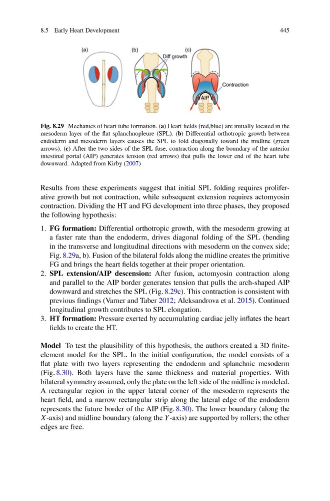
Fig. 8.29 Mechanics of heart tube formation. (a) Heart fields (red,blue) are initially located in the
mesoderm layer of the flat splanchnopleure (SPL). (b) Differential orthotropic growth between
endoderm and mesoderm layers causes the SPL to fold diagonally toward the midline (green
arrows). (c) After the two sides of the SPL fuse, contraction along the boundary of the anterior
intestinal portal (AIP) generates tension (red arrows) that pulls the lower end of the heart tube
downward. Adapted from Kirby (2007)
Results from these experiments suggest that initial SPL folding requires proliferative growth but not contraction, while subsequent extension requires actomyosin
contraction. Dividing the HT and FG development into three phases, they proposed
the following hypothesis:
1. FG formation: Differential orthotropic growth, with the mesoderm growing at
a faster rate than the endoderm, drives diagonal folding of the SPL (bending
in the transverse and longitudinal directions with mesoderm on the convex side;
Fig. 8.29a, b). Fusion of the bilateral folds along the midline creates the primitive
FG and brings the heart fields together at their proper orientation.
2. SPL extension/AIP descension: After fusion, actomyosin contraction along
and parallel to the AIP border generates tension that pulls the arch-shaped AIP
downward and stretches the SPL (Fig. 8.29c). This contraction is consistent with
previous findings (Varner and Taber 2012; Aleksandrova et al. 2015). Continued
longitudinal growth contributes to SPL elongation.
3. HT formation: Pressure exerted by accumulating cardiac jelly inflates the heart
fields to create the HT.
Model To test the plausibility of this hypothesis, the authors created a 3D finiteelement model for the SPL. In the initial configuration, the model consists of a
flat plate with two layers representing the endoderm and splanchnic mesoderm
(Fig. 8.30). Both layers have the same thickness and material properties. With
bilateral symmetry assumed, only the plate on the left side of the midline is modeled.
A rectangular region in the upper lateral corner of the mesoderm represents the
heart field, and a narrow rectangular strip along the lateral edge of the endoderm
represents the future border of the AIP (Fig. 8.30). The lower boundary (along the
X-axis) and midline boundary (along the Y -axis) are supported by rollers; the other
edges are free.
445
Fig. 8.29 Mechanics of heart tube formation. (a) Heart fields (red,blue) are initially located in the
mesoderm layer of the flat splanchnopleure (SPL). (b) Differential orthotropic growth between
endoderm and mesoderm layers causes the SPL to fold diagonally toward the midline (green
arrows). (c) After the two sides of the SPL fuse, contraction along the boundary of the anterior
intestinal portal (AIP) generates tension (red arrows) that pulls the lower end of the heart tube
downward. Adapted from Kirby (2007)
Results from these experiments suggest that initial SPL folding requires proliferative growth but not contraction, while subsequent extension requires actomyosin
contraction. Dividing the HT and FG development into three phases, they proposed
the following hypothesis:
1. FG formation: Differential orthotropic growth, with the mesoderm growing at
a faster rate than the endoderm, drives diagonal folding of the SPL (bending
in the transverse and longitudinal directions with mesoderm on the convex side;
Fig. 8.29a, b). Fusion of the bilateral folds along the midline creates the primitive
FG and brings the heart fields together at their proper orientation.
2. SPL extension/AIP descension: After fusion, actomyosin contraction along
and parallel to the AIP border generates tension that pulls the arch-shaped AIP
downward and stretches the SPL (Fig. 8.29c). This contraction is consistent with
previous findings (Varner and Taber 2012; Aleksandrova et al. 2015). Continued
longitudinal growth contributes to SPL elongation.
3. HT formation: Pressure exerted by accumulating cardiac jelly inflates the heart
fields to create the HT.
Model To test the plausibility of this hypothesis, the authors created a 3D finiteelement model for the SPL. In the initial configuration, the model consists of a
flat plate with two layers representing the endoderm and splanchnic mesoderm
(Fig. 8.30). Both layers have the same thickness and material properties. With
bilateral symmetry assumed, only the plate on the left side of the midline is modeled.
A rectangular region in the upper lateral corner of the mesoderm represents the
heart field, and a narrow rectangular strip along the lateral edge of the endoderm
represents the future border of the AIP (Fig. 8.30). The lower boundary (along the
X-axis) and midline boundary (along the Y -axis) are supported by rollers; the other
edges are free.