

8.5 Early Heart Development
443
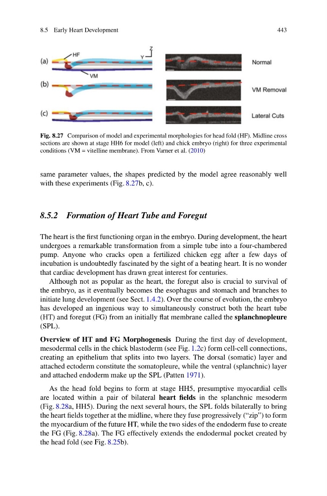
Fig. 8.27 Comparison of model and experimental morphologies for head fold (HF). Midline cross
sections are shown at stage HH6 for model (left) and chick embryo (right) for three experimental
conditions (VM = vitelline membrane). From Varner et al. (2010)
same parameter values, the shapes predicted by the model agree reasonably well
with these experiments (Fig. 8.27b, c).
8.5.2 Formation of Heart Tube and Foregut
The heart is the first functioning organ in the embryo. During development, the heart
undergoes a remarkable transformation from a simple tube into a four-chambered
pump. Anyone who cracks open a fertilized chicken egg after a few days of
incubation is undoubtedly fascinated by the sight of a beating heart. It is no wonder
that cardiac development has drawn great interest for centuries.
Although not as popular as the heart, the foregut also is crucial to survival of
the embryo, as it eventually becomes the esophagus and stomach and branches to
initiate lung development (see Sect. 1.4.2). Over the course of evolution, the embryo
has developed an ingenious way to simultaneously construct both the heart tube
(HT) and foregut (FG) from an initially flat membrane called the splanchnopleure
(SPL).
Overview of HT and FG Morphogenesis During the first day of development,
mesodermal cells in the chick blastoderm (see Fig. 1.2c) form cell-cell connections,
creating an epithelium that splits into two layers. The dorsal (somatic) layer and
attached ectoderm constitute the somatopleure, while the ventral (splanchnic) layer
and attached endoderm make up the SPL (Patten 1971).
As the head fold begins to form at stage HH5, presumptive myocardial cells
are located within a pair of bilateral heart fields in the splanchnic mesoderm
(Fig. 8.28a, HH5). During the next several hours, the SPL folds bilaterally to bring
the heart fields together at the midline, where they fuse progressively (“zip”) to form
the myocardium of the future HT, while the two sides of the endoderm fuse to create
the FG (Fig. 8.28a). The FG effectively extends the endodermal pocket created by
the head fold (see Fig. 8.25b).
443
Fig. 8.27 Comparison of model and experimental morphologies for head fold (HF). Midline cross
sections are shown at stage HH6 for model (left) and chick embryo (right) for three experimental
conditions (VM = vitelline membrane). From Varner et al. (2010)
same parameter values, the shapes predicted by the model agree reasonably well
with these experiments (Fig. 8.27b, c).
8.5.2 Formation of Heart Tube and Foregut
The heart is the first functioning organ in the embryo. During development, the heart
undergoes a remarkable transformation from a simple tube into a four-chambered
pump. Anyone who cracks open a fertilized chicken egg after a few days of
incubation is undoubtedly fascinated by the sight of a beating heart. It is no wonder
that cardiac development has drawn great interest for centuries.
Although not as popular as the heart, the foregut also is crucial to survival of
the embryo, as it eventually becomes the esophagus and stomach and branches to
initiate lung development (see Sect. 1.4.2). Over the course of evolution, the embryo
has developed an ingenious way to simultaneously construct both the heart tube
(HT) and foregut (FG) from an initially flat membrane called the splanchnopleure
(SPL).
Overview of HT and FG Morphogenesis During the first day of development,
mesodermal cells in the chick blastoderm (see Fig. 1.2c) form cell-cell connections,
creating an epithelium that splits into two layers. The dorsal (somatic) layer and
attached ectoderm constitute the somatopleure, while the ventral (splanchnic) layer
and attached endoderm make up the SPL (Patten 1971).
As the head fold begins to form at stage HH5, presumptive myocardial cells
are located within a pair of bilateral heart fields in the splanchnic mesoderm
(Fig. 8.28a, HH5). During the next several hours, the SPL folds bilaterally to bring
the heart fields together at the midline, where they fuse progressively (“zip”) to form
the myocardium of the future HT, while the two sides of the endoderm fuse to create
the FG (Fig. 8.28a). The FG effectively extends the endodermal pocket created by
the head fold (see Fig. 8.25b).