

436
8 Morphogenesis
also speculated that cytoplasm displaced from the shrinking apical side forces
the basal side to expand, enhancing the change in cell shape from columnar to
trapezoidal. During the final 25% of gastrulation, this mechanism is simulated here
by radial contraction ( ˙
G r < 0) and circumferential expansion (G θ = 1/G r for
incompressible cytoplasm) in the mesoderm. The model then produces a closed
tube (Fig. 8.19d), similar to the actual morphology (Fig. 8.18b, right). This potential
mechanism and the cause of mesodermal thinning warrant further study.
8.4.3 Gastrulation in Sea Urchins
The sea urchin blastula is a fluid-filled spherical shell. Gastrulation consists of
two phases (Keller et al. 2003). During primary invagination, a relatively shallow,
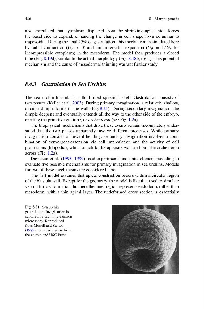
circular dimple forms in the wall (Fig. 8.21). During secondary invagination, the
dimple deepens and eventually extends all the way to the other side of the embryo,
creating the primitive gut tube, or archenteron (see Fig. 1.2a).
The biophysical mechanisms that drive these events remain incompletely understood, but the two phases apparently involve different processes. While primary
invagination consists of inward bending, secondary invagination involves a combination of convergent-extension via cell intercalation and the activity of cell
protrusions (filopodia), which attach to the opposite wall and pull the archenteron
across (Fig. 1.2a).
Davidson et al. (1995, 1999) used experiments and finite-element modeling to
evaluate five possible mechanisms for primary invagination in sea urchins. Models
for two of these mechanisms are considered here.
The first model assumes that apical constriction occurs within a circular region
of the blastula wall. Except for the geometry, the model is like that used to simulate
ventral furrow formation, but here the inner region represents endoderm, rather than
mesoderm, with a thin apical layer. The undeformed cross section is essentially
Fig. 8.21 Sea urchin
gastrulation. Invagination is
captured by scanning electron
microscopy. Reproduced
from Morrill and Santos
(1985), with permission from
the editors and USC Press
8 Morphogenesis
also speculated that cytoplasm displaced from the shrinking apical side forces
the basal side to expand, enhancing the change in cell shape from columnar to
trapezoidal. During the final 25% of gastrulation, this mechanism is simulated here
by radial contraction ( ˙
G r < 0) and circumferential expansion (G θ = 1/G r for
incompressible cytoplasm) in the mesoderm. The model then produces a closed
tube (Fig. 8.19d), similar to the actual morphology (Fig. 8.18b, right). This potential
mechanism and the cause of mesodermal thinning warrant further study.
8.4.3 Gastrulation in Sea Urchins
The sea urchin blastula is a fluid-filled spherical shell. Gastrulation consists of
two phases (Keller et al. 2003). During primary invagination, a relatively shallow,
circular dimple forms in the wall (Fig. 8.21). During secondary invagination, the
dimple deepens and eventually extends all the way to the other side of the embryo,
creating the primitive gut tube, or archenteron (see Fig. 1.2a).
The biophysical mechanisms that drive these events remain incompletely understood, but the two phases apparently involve different processes. While primary
invagination consists of inward bending, secondary invagination involves a combination of convergent-extension via cell intercalation and the activity of cell
protrusions (filopodia), which attach to the opposite wall and pull the archenteron
across (Fig. 1.2a).
Davidson et al. (1995, 1999) used experiments and finite-element modeling to
evaluate five possible mechanisms for primary invagination in sea urchins. Models
for two of these mechanisms are considered here.
The first model assumes that apical constriction occurs within a circular region
of the blastula wall. Except for the geometry, the model is like that used to simulate
ventral furrow formation, but here the inner region represents endoderm, rather than
mesoderm, with a thin apical layer. The undeformed cross section is essentially
Fig. 8.21 Sea urchin
gastrulation. Invagination is
captured by scanning electron
microscopy. Reproduced
from Morrill and Santos
(1985), with permission from
the editors and USC Press