

8.4 Gastrulation
435
layer (apex) and a thick inner layer (mesoderm). Contraction is simulated through
the growth tensor
G = G r e r e r + G θ e θ e θ + e z e z ,
where the e i are cylindrical base vectors in the undeformed configuration. This
model includes no external constraints.
First, consider the case where the apex undergoes circumferential contraction
(G θ < 1, G r = 1 with 10x modulus), while the rest of the model remains passive.
For G θ = 0.3, the shape of the invagination is rather broad, and the sides of the
model bow outward (Fig. 8.19b). These results are in rather poor agreement with
observations (Fig. 8.18b).
Including other factors can generate more realistic morphologies. For example, it
is known that yolk squirts out when a Drosophila embryo is punctured (Conte et al.
2008), suggesting that internal pressure may be important. Applying pressure to the
inner surface pushes the invaginating region upward, giving a much more realistic
shape for G θ = 0.05 (Fig. 8.19c). However, the invagination remains open, contrary
to observations.
To prevent outward bulging, some researchers include the vitelline membrane as
a rigid, frictionless shell surrounding the embryo (Munoz et al. 2007; Conte et al.
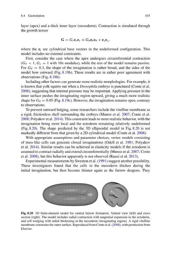
2008; Polyakov et al. 2014). This constraint leads to more realistic behavior, with the
invagination being more local and the ectoderm remaining relatively undeformed
(Fig. 8.20). The shape predicted by the 3D ellipsoidal model in Fig. 8.20 is not
markedly different from that given by a 2D cylindrical model (Conte et al. 2008).
With appropriate assumptions and parameter choices, vertex models consisting
of truss-like cells can generate closed invaginations (Odell et al. 1981; Polyakov
et al. 2014). Similar results can be achieved in elasticity models if the ectoderm is
assumed to contract radially and extend circumferentially (Munoz et al. 2007; Conte
et al. 2008), but this behavior apparently is not observed (Rauzi et al. 2013).
Experimental measurements by Sweeton et al. (1991) suggest another possibility.
These investigators found that the cells in the mesoderm thicken during the
initial invagination, but then become thinner again as the furrow deepens. They
Fig. 8.20 3D finite-element model for ventral furrow formation. Ventral view (left) and cross
section (right). The model includes radial contraction with tangential expansion in the ectoderm,
and cell wedging with radial thickening in the mesoderm (invaginating region). A rigid vitelline
membrane constrains the outer surface. Reproduced from Conte et al. (2008), with permission from
Elsevier
435
layer (apex) and a thick inner layer (mesoderm). Contraction is simulated through
the growth tensor
G = G r e r e r + G θ e θ e θ + e z e z ,
where the e i are cylindrical base vectors in the undeformed configuration. This
model includes no external constraints.
First, consider the case where the apex undergoes circumferential contraction
(G θ < 1, G r = 1 with 10x modulus), while the rest of the model remains passive.
For G θ = 0.3, the shape of the invagination is rather broad, and the sides of the
model bow outward (Fig. 8.19b). These results are in rather poor agreement with
observations (Fig. 8.18b).
Including other factors can generate more realistic morphologies. For example, it
is known that yolk squirts out when a Drosophila embryo is punctured (Conte et al.
2008), suggesting that internal pressure may be important. Applying pressure to the
inner surface pushes the invaginating region upward, giving a much more realistic
shape for G θ = 0.05 (Fig. 8.19c). However, the invagination remains open, contrary
to observations.
To prevent outward bulging, some researchers include the vitelline membrane as
a rigid, frictionless shell surrounding the embryo (Munoz et al. 2007; Conte et al.
2008; Polyakov et al. 2014). This constraint leads to more realistic behavior, with the
invagination being more local and the ectoderm remaining relatively undeformed
(Fig. 8.20). The shape predicted by the 3D ellipsoidal model in Fig. 8.20 is not
markedly different from that given by a 2D cylindrical model (Conte et al. 2008).
With appropriate assumptions and parameter choices, vertex models consisting
of truss-like cells can generate closed invaginations (Odell et al. 1981; Polyakov
et al. 2014). Similar results can be achieved in elasticity models if the ectoderm is
assumed to contract radially and extend circumferentially (Munoz et al. 2007; Conte
et al. 2008), but this behavior apparently is not observed (Rauzi et al. 2013).
Experimental measurements by Sweeton et al. (1991) suggest another possibility.
These investigators found that the cells in the mesoderm thicken during the
initial invagination, but then become thinner again as the furrow deepens. They
Fig. 8.20 3D finite-element model for ventral furrow formation. Ventral view (left) and cross
section (right). The model includes radial contraction with tangential expansion in the ectoderm,
and cell wedging with radial thickening in the mesoderm (invaginating region). A rigid vitelline
membrane constrains the outer surface. Reproduced from Conte et al. (2008), with permission from
Elsevier