

380
7 Remodeling
P
r
θ
φ
P
)
b
(
)
a
(
(c)
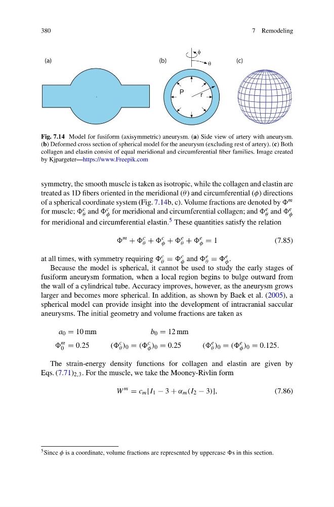
Fig. 7.14 Model for fusiform (axisymmetric) aneurysm. (a) Side view of artery with aneurysm.
(b) Deformed cross section of spherical model for the aneurysm (excluding rest of artery). (c) Both
collagen and elastin consist of equal meridional and circumferential fiber families. Image created
by Kjpargeter—https://www.Freepik.com
symmetry, the smooth muscle is taken as isotropic, while the collagen and elastin are
treated as 1D fibers oriented in the meridional (θ ) and circumferential (φ) directions
of a spherical coordinate system (Fig. 7.14b, c). Volume fractions are denoted by m
for muscle; c
θ and c
φ for meridional and circumferential collagen; and e
θ and e
φ
for meridional and circumferential elastin. 5 These quantities satisfy the relation
m
+
c
θ +
c
φ +
e
θ +
e
φ = 1
(7.85)
at all times, with symmetry requiring c
θ = c
φ and e
θ = e
φ .
Because the model is spherical, it cannot be used to study the early stages of
fusiform aneurysm formation, when a local region begins to bulge outward from
the wall of a cylindrical tube. Accuracy improves, however, as the aneurysm grows
larger and becomes more spherical. In addition, as shown by Baek et al. (2005), a
spherical model can provide insight into the development of intracranial saccular
aneurysms. The initial geometry and volume fractions are taken as
a 0 = 10 mm
b 0 = 12 mm
m
0 = 0.25
((
c
θ ) 0 = ((
c
φ ) 0 = 0.25
((
e
θ ) 0 = ((
e
φ ) 0 = 0.125.
The strain-energy density functions for collagen and elastin are given by
Eqs. (7.71) 2,3 . For the muscle, we take the Mooney-Rivlin form
W
m
= c m [I 1 − 3 + α m (I 2 − 3)],
(7.86)
5 Since φ is a coordinate, volume fractions are represented by uppercase s in this section.
7 Remodeling
P
r
θ
φ
P
)
b
(
)
a
(
(c)
Fig. 7.14 Model for fusiform (axisymmetric) aneurysm. (a) Side view of artery with aneurysm.
(b) Deformed cross section of spherical model for the aneurysm (excluding rest of artery). (c) Both
collagen and elastin consist of equal meridional and circumferential fiber families. Image created
by Kjpargeter—https://www.Freepik.com
symmetry, the smooth muscle is taken as isotropic, while the collagen and elastin are
treated as 1D fibers oriented in the meridional (θ ) and circumferential (φ) directions
of a spherical coordinate system (Fig. 7.14b, c). Volume fractions are denoted by m
for muscle; c
θ and c
φ for meridional and circumferential collagen; and e
θ and e
φ
for meridional and circumferential elastin. 5 These quantities satisfy the relation
m
+
c
θ +
c
φ +
e
θ +
e
φ = 1
(7.85)
at all times, with symmetry requiring c
θ = c
φ and e
θ = e
φ .
Because the model is spherical, it cannot be used to study the early stages of
fusiform aneurysm formation, when a local region begins to bulge outward from
the wall of a cylindrical tube. Accuracy improves, however, as the aneurysm grows
larger and becomes more spherical. In addition, as shown by Baek et al. (2005), a
spherical model can provide insight into the development of intracranial saccular
aneurysms. The initial geometry and volume fractions are taken as
a 0 = 10 mm
b 0 = 12 mm
m
0 = 0.25
((
c
θ ) 0 = ((
c
φ ) 0 = 0.25
((
e
θ ) 0 = ((
e
φ ) 0 = 0.125.
The strain-energy density functions for collagen and elastin are given by
Eqs. (7.71) 2,3 . For the muscle, we take the Mooney-Rivlin form
W
m
= c m [I 1 − 3 + α m (I 2 − 3)],
(7.86)
5 Since φ is a coordinate, volume fractions are represented by uppercase s in this section.