

7.6 Case Study: Growth and Remodeling of Arteries
379
7.6.2 Aneurysm Development
An aneurysm is a bulge that forms in the wall of the heart or an artery. Multiple
factors can cause an aneurysm, including local loss of elastin and smooth muscle
tone. By Laplace’s law, wall stress increases with the radius of the bulge. Studies in
arteries have shown that elevated wall stress increases the risk of rupture (Fillinger
et al. 2002, 2003), which usually proves fatal.
In judging whether to intervene surgically, physicians generally use aneurysm
size as a relatively crude indicator of rupture potential. This approach is not always
reliable, however, as two aneurysms with the same size and location can progress
differently, with one continuing to grow and eventually rupture and the other
stabilizing and causing no major problems. Thus, it is important to understand the
role that mechanics plays in the formation, expansion, and rupture of aneurysms.
This section focuses on vascular aneurysms.
Arterial aneurysms form most often in the brain and aorta, with their shape
depending on location. Fusiform aneurysms are symmetric relative to the centerline
of a blood vessel, whereas saccular aneurysms are asymmetric, bulging to one
side. Since wall stress depends on geometry, researchers have devoted considerable
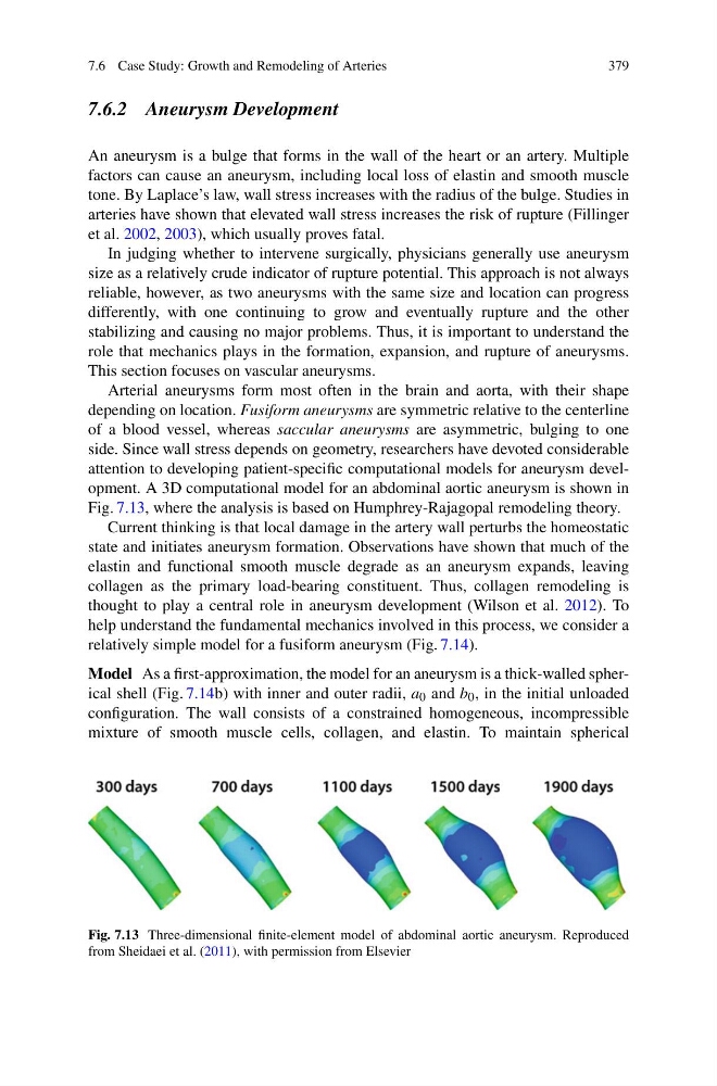
attention to developing patient-specific computational models for aneurysm development. A 3D computational model for an abdominal aortic aneurysm is shown in
Fig. 7.13, where the analysis is based on Humphrey-Rajagopal remodeling theory.
Current thinking is that local damage in the artery wall perturbs the homeostatic
state and initiates aneurysm formation. Observations have shown that much of the
elastin and functional smooth muscle degrade as an aneurysm expands, leaving
collagen as the primary load-bearing constituent. Thus, collagen remodeling is
thought to play a central role in aneurysm development (Wilson et al. 2012). To
help understand the fundamental mechanics involved in this process, we consider a
relatively simple model for a fusiform aneurysm (Fig. 7.14).
Model As a first-approximation, the model for an aneurysm is a thick-walled spherical shell (Fig. 7.14b) with inner and outer radii, a 0 and b 0 , in the initial unloaded
configuration. The wall consists of a constrained homogeneous, incompressible
mixture of smooth muscle cells, collagen, and elastin. To maintain spherical
Fig. 7.13 Three-dimensional finite-element model of abdominal aortic aneurysm. Reproduced
from Sheidaei et al. (2011), with permission from Elsevier
379
7.6.2 Aneurysm Development
An aneurysm is a bulge that forms in the wall of the heart or an artery. Multiple
factors can cause an aneurysm, including local loss of elastin and smooth muscle
tone. By Laplace’s law, wall stress increases with the radius of the bulge. Studies in
arteries have shown that elevated wall stress increases the risk of rupture (Fillinger
et al. 2002, 2003), which usually proves fatal.
In judging whether to intervene surgically, physicians generally use aneurysm
size as a relatively crude indicator of rupture potential. This approach is not always
reliable, however, as two aneurysms with the same size and location can progress
differently, with one continuing to grow and eventually rupture and the other
stabilizing and causing no major problems. Thus, it is important to understand the
role that mechanics plays in the formation, expansion, and rupture of aneurysms.
This section focuses on vascular aneurysms.
Arterial aneurysms form most often in the brain and aorta, with their shape
depending on location. Fusiform aneurysms are symmetric relative to the centerline
of a blood vessel, whereas saccular aneurysms are asymmetric, bulging to one
side. Since wall stress depends on geometry, researchers have devoted considerable
attention to developing patient-specific computational models for aneurysm development. A 3D computational model for an abdominal aortic aneurysm is shown in
Fig. 7.13, where the analysis is based on Humphrey-Rajagopal remodeling theory.
Current thinking is that local damage in the artery wall perturbs the homeostatic
state and initiates aneurysm formation. Observations have shown that much of the
elastin and functional smooth muscle degrade as an aneurysm expands, leaving
collagen as the primary load-bearing constituent. Thus, collagen remodeling is
thought to play a central role in aneurysm development (Wilson et al. 2012). To
help understand the fundamental mechanics involved in this process, we consider a
relatively simple model for a fusiform aneurysm (Fig. 7.14).
Model As a first-approximation, the model for an aneurysm is a thick-walled spherical shell (Fig. 7.14b) with inner and outer radii, a 0 and b 0 , in the initial unloaded
configuration. The wall consists of a constrained homogeneous, incompressible
mixture of smooth muscle cells, collagen, and elastin. To maintain spherical
Fig. 7.13 Three-dimensional finite-element model of abdominal aortic aneurysm. Reproduced
from Sheidaei et al. (2011), with permission from Elsevier