

330
6 Growth
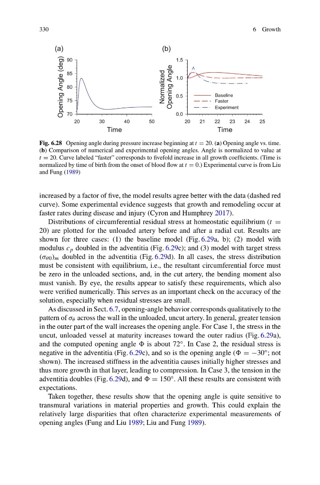
Fig. 6.28 Opening angle during pressure increase beginning at t = 20. (a) Opening angle vs. time.
(b) Comparison of numerical and experimental opening angles. Angle is normalized to value at
t = 20. Curve labeled “faster” corresponds to fivefold increase in all growth coefficients. (Time is
normalized by time of birth from the onset of blood flow at t = 0.) Experimental curve is from Liu
and Fung (1989)
increased by a factor of five, the model results agree better with the data (dashed red
curve). Some experimental evidence suggests that growth and remodeling occur at
faster rates during disease and injury (Cyron and Humphrey 2017).
Distributions of circumferential residual stress at homeostatic equilibrium (t =
20) are plotted for the unloaded artery before and after a radial cut. Results are
shown for three cases: (1) the baseline model (Fig. 6.29a, b); (2) model with
modulus c p doubled in the adventitia (Fig. 6.29c); and (3) model with target stress
(σ θ0 ) m doubled in the adventitia (Fig. 6.29d). In all cases, the stress distribution
must be consistent with equilibrium, i.e., the resultant circumferential force must
be zero in the unloaded sections, and, in the cut artery, the bending moment also
must vanish. By eye, the results appear to satisfy these requirements, which also
were verified numerically. This serves as an important check on the accuracy of the
solution, especially when residual stresses are small.
As discussed in Sect. 6.7, opening-angle behavior corresponds qualitatively to the
pattern of σ θ across the wall in the unloaded, uncut artery. In general, greater tension
in the outer part of the wall increases the opening angle. For Case 1, the stress in the
uncut, unloaded vessel at maturity increases toward the outer radius (Fig. 6.29a),
and the computed opening angle is about 72 ◦ . In Case 2, the residual stress is
negative in the adventitia (Fig. 6.29c), and so is the opening angle ( = −30 ◦ ; not
shown). The increased stiffness in the adventitia causes initially higher stresses and
thus more growth in that layer, leading to compression. In Case 3, the tension in the
adventitia doubles (Fig. 6.29d), and = 150 ◦ . All these results are consistent with
expectations.
Taken together, these results show that the opening angle is quite sensitive to
transmural variations in material properties and growth. This could explain the
relatively large disparities that often characterize experimental measurements of
opening angles (Fung and Liu 1989; Liu and Fung 1989).
6 Growth
Fig. 6.28 Opening angle during pressure increase beginning at t = 20. (a) Opening angle vs. time.
(b) Comparison of numerical and experimental opening angles. Angle is normalized to value at
t = 20. Curve labeled “faster” corresponds to fivefold increase in all growth coefficients. (Time is
normalized by time of birth from the onset of blood flow at t = 0.) Experimental curve is from Liu
and Fung (1989)
increased by a factor of five, the model results agree better with the data (dashed red
curve). Some experimental evidence suggests that growth and remodeling occur at
faster rates during disease and injury (Cyron and Humphrey 2017).
Distributions of circumferential residual stress at homeostatic equilibrium (t =
20) are plotted for the unloaded artery before and after a radial cut. Results are
shown for three cases: (1) the baseline model (Fig. 6.29a, b); (2) model with
modulus c p doubled in the adventitia (Fig. 6.29c); and (3) model with target stress
(σ θ0 ) m doubled in the adventitia (Fig. 6.29d). In all cases, the stress distribution
must be consistent with equilibrium, i.e., the resultant circumferential force must
be zero in the unloaded sections, and, in the cut artery, the bending moment also
must vanish. By eye, the results appear to satisfy these requirements, which also
were verified numerically. This serves as an important check on the accuracy of the
solution, especially when residual stresses are small.
As discussed in Sect. 6.7, opening-angle behavior corresponds qualitatively to the
pattern of σ θ across the wall in the unloaded, uncut artery. In general, greater tension
in the outer part of the wall increases the opening angle. For Case 1, the stress in the
uncut, unloaded vessel at maturity increases toward the outer radius (Fig. 6.29a),
and the computed opening angle is about 72 ◦ . In Case 2, the residual stress is
negative in the adventitia (Fig. 6.29c), and so is the opening angle ( = −30 ◦ ; not
shown). The increased stiffness in the adventitia causes initially higher stresses and
thus more growth in that layer, leading to compression. In Case 3, the tension in the
adventitia doubles (Fig. 6.29d), and = 150 ◦ . All these results are consistent with
expectations.
Taken together, these results show that the opening angle is quite sensitive to
transmural variations in material properties and growth. This could explain the
relatively large disparities that often characterize experimental measurements of
opening angles (Fung and Liu 1989; Liu and Fung 1989).