

27
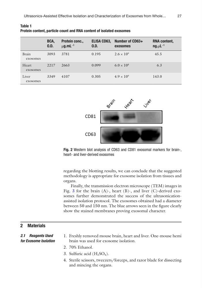
regarding the blotting results, we can conclude that the suggested
methodology is appropriate for exosome isolation from tissues and
organs.
Finally, the transmission electron microscope (TEM) images in
Fig. 3 for the brain (A)-, heart (B)-, and liver (C)-derived exosomes further demonstrated the success of the ultrasonicationassisted isolation protocol. The exosomes obtained had a diameter
between 50 and 150 nm. The blue arrows seen in the figure clearly
show the stained membranes proving exosomal character.
2 Materials
1. Freshly removed mouse brain, heart and liver. One-mouse hemi
brain was used for exosome isolation.
2. 70% Ethanol.
3. Sulfuric acid (H 2 SO 4 ).
4. Sterile scissors, tweezers/forceps, and razor blade for dissecting
and mincing the organs.
2.1 Reagents Used
for Exosome Isolation
Table 1
Protein content, particle count and RNA content of isolated exosomes
BCA,
O.D.
Protein conc.,
μg.mL
−1
ELISA CD63,
O.D.
Number of CD63+
exosomes
RNA content,
ng.μL
−1
Brain
exosomes
3093
3781
0.195
2.6 × 10
9
45.5
Heart
exosomes
2217
2663
0.099
6.0 × 10
8
6.3
Liver
exosomes
3349
4107
0.305
4.9 × 10
9
143.0
Fig. 2 Western blot analysis of CD63 and CD81 exosomal markers for brain-,
heart- and liver-derived exosomes
Ultrasonics-Assisted Effective Isolation and Characterization of Exosomes from Whole…
regarding the blotting results, we can conclude that the suggested
methodology is appropriate for exosome isolation from tissues and
organs.
Finally, the transmission electron microscope (TEM) images in
Fig. 3 for the brain (A)-, heart (B)-, and liver (C)-derived exosomes further demonstrated the success of the ultrasonicationassisted isolation protocol. The exosomes obtained had a diameter
between 50 and 150 nm. The blue arrows seen in the figure clearly
show the stained membranes proving exosomal character.
2 Materials
1. Freshly removed mouse brain, heart and liver. One-mouse hemi
brain was used for exosome isolation.
2. 70% Ethanol.
3. Sulfuric acid (H 2 SO 4 ).
4. Sterile scissors, tweezers/forceps, and razor blade for dissecting
and mincing the organs.
2.1 Reagents Used
for Exosome Isolation
Table 1
Protein content, particle count and RNA content of isolated exosomes
BCA,
O.D.
Protein conc.,
μg.mL
−1
ELISA CD63,
O.D.
Number of CD63+
exosomes
RNA content,
ng.μL
−1
Brain
exosomes
3093
3781
0.195
2.6 × 10
9
45.5
Heart
exosomes
2217
2663
0.099
6.0 × 10
8
6.3
Liver
exosomes
3349
4107
0.305
4.9 × 10
9
143.0
Fig. 2 Western blot analysis of CD63 and CD81 exosomal markers for brain-,
heart- and liver-derived exosomes
Ultrasonics-Assisted Effective Isolation and Characterization of Exosomes from Whole…