

28
5. Sterile and cold phosphate-buffered saline (1× PBS, pH 7.4).
6. 50 mL Conical tubes.
7. Polyethylene glycol 4000 (PEG 4000).
8. 0.2 μm Filter adapters with surfactant-free cellulose acetate
membrane and 10 mL plunge syringes.
1. 1× RIPA lysis buffer (1× lysis buffer, 200 mM PMSF, inhibitor
cocktail, and sodium orthovanadate) (Santa Cruz sc-24948).
2. BCA kit (Thermo Scientific 23227).
3. Flat-bottom 96-well microplate and adhesive covers.
4. CD63 and CD81 ELISA kits (System Bioscience EXOELCD63A- 1 and EXOEL-CD81A-1).
1. Acrylamide/bis-acrylamide, 30% solution (Sigma).
2. Tris (pH 8.8, 0.5 M).
2.2 Reagents Used
for the
Characterization
of Isolated Exosomes
2.2.1 Exosomal Protein
Quantification
2.2.2 Western Blot
Analysis
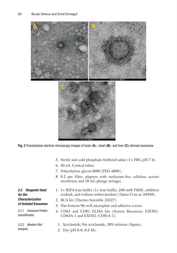
Fig. 3 Transmission electron microscopy images of brain (A)-, heart (B)- and liver (C)-derived exosomes
Burak Derkus and Emel Emregul
5. Sterile and cold phosphate-buffered saline (1× PBS, pH 7.4).
6. 50 mL Conical tubes.
7. Polyethylene glycol 4000 (PEG 4000).
8. 0.2 μm Filter adapters with surfactant-free cellulose acetate
membrane and 10 mL plunge syringes.
1. 1× RIPA lysis buffer (1× lysis buffer, 200 mM PMSF, inhibitor
cocktail, and sodium orthovanadate) (Santa Cruz sc-24948).
2. BCA kit (Thermo Scientific 23227).
3. Flat-bottom 96-well microplate and adhesive covers.
4. CD63 and CD81 ELISA kits (System Bioscience EXOELCD63A- 1 and EXOEL-CD81A-1).
1. Acrylamide/bis-acrylamide, 30% solution (Sigma).
2. Tris (pH 8.8, 0.5 M).
2.2 Reagents Used
for the
Characterization
of Isolated Exosomes
2.2.1 Exosomal Protein
Quantification
2.2.2 Western Blot
Analysis
Fig. 3 Transmission electron microscopy images of brain (A)-, heart (B)- and liver (C)-derived exosomes
Burak Derkus and Emel Emregul