

26
between cell lines [10]. Moreover, the cellular content does not
fully reflect the molecular composition of tissues. Since the exosomes isolated from secondary cell lines do not fully reflect the
properties of the primary cells or original tissue [11], it is difficult
to judge the molecular contents of exosomes. It is important to
know the exact molecular content of the exosomes during secretion from the tissue so that the diagnostic and therapeutic
approaches can be properly examined. In addition, in order to fully
illuminate the mechanisms of exosomes in cellular communication,
attempted in various publications [12], isolation from natural environments namely living tissues and organs would be preferred
instead of cell culture CM. However, it can be seen in the literature
that there are only a couple studies dealing with the isolation of
exosomes from whole tissues/organs [13]. This study mainly
focuses on the effective isolation of exosomes from heart, brain
and liver. Ultrasonication was for the first time applied in this study
for exosome isolation that loosened the extracellular space of
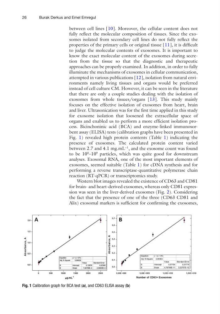
organs and enabled us to perform a more efficient isolation process. Bicinchoninic acid (BCA) and enzyme-linked immunosorbent assay (ELISA) tests (calibration graphs have been presented in
Fig. 1) revealed high protein contents (Table 1) indicating the
presence of exosomes. The calculated protein content varied
between 2.7 and 4.1 mg.mL
−1
, and the exosome count was found
to be 10
8
–10
9
particles, which was quite good for downstream
analyses. Exosomal RNA, one of the most important elements of
exosomes, seemed suitable (Table 1) for cDNA synthesis and for
performing a reverse transcriptase-quantitative polymerase chain
reaction (RT-qPCR) or transcriptomics study.
Western blot images revealed the existence of CD63 and CD81
for brain- and heart-derived exosomes, whereas only CD81 expression was seen in the liver-derived exosomes (Fig. 2). Considering
the fact that the presence of one of the three (CD63 CD81 and
Alix) exosomal markers is sufficient for confirming the exosomes,
Fig. 1 Calibration graph for BCA test (a), and CD63 ELISA assay (b)
Burak Derkus and Emel Emregul
between cell lines [10]. Moreover, the cellular content does not
fully reflect the molecular composition of tissues. Since the exosomes isolated from secondary cell lines do not fully reflect the
properties of the primary cells or original tissue [11], it is difficult
to judge the molecular contents of exosomes. It is important to
know the exact molecular content of the exosomes during secretion from the tissue so that the diagnostic and therapeutic
approaches can be properly examined. In addition, in order to fully
illuminate the mechanisms of exosomes in cellular communication,
attempted in various publications [12], isolation from natural environments namely living tissues and organs would be preferred
instead of cell culture CM. However, it can be seen in the literature
that there are only a couple studies dealing with the isolation of
exosomes from whole tissues/organs [13]. This study mainly
focuses on the effective isolation of exosomes from heart, brain
and liver. Ultrasonication was for the first time applied in this study
for exosome isolation that loosened the extracellular space of
organs and enabled us to perform a more efficient isolation process. Bicinchoninic acid (BCA) and enzyme-linked immunosorbent assay (ELISA) tests (calibration graphs have been presented in
Fig. 1) revealed high protein contents (Table 1) indicating the
presence of exosomes. The calculated protein content varied
between 2.7 and 4.1 mg.mL
−1
, and the exosome count was found
to be 10
8
–10
9
particles, which was quite good for downstream
analyses. Exosomal RNA, one of the most important elements of
exosomes, seemed suitable (Table 1) for cDNA synthesis and for
performing a reverse transcriptase-quantitative polymerase chain
reaction (RT-qPCR) or transcriptomics study.
Western blot images revealed the existence of CD63 and CD81
for brain- and heart-derived exosomes, whereas only CD81 expression was seen in the liver-derived exosomes (Fig. 2). Considering
the fact that the presence of one of the three (CD63 CD81 and
Alix) exosomal markers is sufficient for confirming the exosomes,
Fig. 1 Calibration graph for BCA test (a), and CD63 ELISA assay (b)
Burak Derkus and Emel Emregul