

95
9. You can perform quantitative analysis to calculate the percentage of wound closure by the equation [24]
Wound closure%
/
,
A
A
A
t h
t xh
t h
0
0
100
where A is the wound area at different time points t = 0 h and xh.
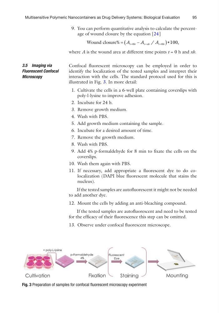
Confocal fluorescent microscopy can be employed in order to
identify the localization of the tested samples and interpret their
interaction with the cells. The standard protocol used for this is
illustrated in Fig. 3. In more detail:
1. Cultivate the cells in a 6-well plate containing coverslips with
poly-l-lysine to improve adhesion.
2. Incubate for 24 h.
3. Remove growth medium.
4. Wash with PBS.
5. Add growth medium containing the sample.
6. Incubate for a desired amount of time.
7. Remove the growth medium.
8. Wash with PBS.
9. Add 4% p-formaldehyde for 8 min to fixate the cells on the
coverslips.
10. Wash them again with PBS.
11. If necessary, add appropriate a fluorescent dye to do colocalization (DAPI blue fluorescent molecule that stains the
nucleus).
If the tested samples are autofluorescent it might not be needed
to add another dye.
12. Mount the cells by adding an anti-bleaching compound.
If the tested samples are autofluorescent and need to be tested
for the efficacy of their fluorescence this step can be omitted.
13. Observe under confocal fluorescent microscope.
3.5 Imaging via
Fluorescent Confocal
Microscopy
Fig. 3 Preparation of samples for confocal fluorescent microscopy experiment
Multisensitive Polymeric Nanocontainers as Drug Delivery Systems: Biological Evaluation
9. You can perform quantitative analysis to calculate the percentage of wound closure by the equation [24]
Wound closure%
/
,
A
A
A
t h
t xh
t h
0
0
100
where A is the wound area at different time points t = 0 h and xh.
Confocal fluorescent microscopy can be employed in order to
identify the localization of the tested samples and interpret their
interaction with the cells. The standard protocol used for this is
illustrated in Fig. 3. In more detail:
1. Cultivate the cells in a 6-well plate containing coverslips with
poly-l-lysine to improve adhesion.
2. Incubate for 24 h.
3. Remove growth medium.
4. Wash with PBS.
5. Add growth medium containing the sample.
6. Incubate for a desired amount of time.
7. Remove the growth medium.
8. Wash with PBS.
9. Add 4% p-formaldehyde for 8 min to fixate the cells on the
coverslips.
10. Wash them again with PBS.
11. If necessary, add appropriate a fluorescent dye to do colocalization (DAPI blue fluorescent molecule that stains the
nucleus).
If the tested samples are autofluorescent it might not be needed
to add another dye.
12. Mount the cells by adding an anti-bleaching compound.
If the tested samples are autofluorescent and need to be tested
for the efficacy of their fluorescence this step can be omitted.
13. Observe under confocal fluorescent microscope.
3.5 Imaging via
Fluorescent Confocal
Microscopy
Fig. 3 Preparation of samples for confocal fluorescent microscopy experiment
Multisensitive Polymeric Nanocontainers as Drug Delivery Systems: Biological Evaluation